 Movie
Movie Controller

[English] 日本語
 Yorodumi
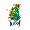
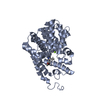
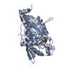
- PDB-1goj: Structure of a fast kinesin: Implications for ATPase mechanism an... -
+ Open data

- Basic information
| Entry | Database: PDB / ID: 1goj | ||||||
|---|---|---|---|---|---|---|---|
| Title | Structure of a fast kinesin: Implications for ATPase mechanism and interactions with microtubules | ||||||
 Components Components | KINESIN HEAVY CHAIN | ||||||
 Keywords Keywords | MOTOR PROTEIN / KINESIN / ATPASE / NEUROSPORA CRASSA | ||||||
| Function / homology |  Function and homology information Function and homology informationcytoskeleton-dependent intracellular transport / plus-end-directed microtubule motor activity / microtubule lateral binding / kinesin complex / microtubule motor activity / microtubule-based movement / microtubule cytoskeleton / microtubule binding / microtubule / ATP hydrolysis activity ...cytoskeleton-dependent intracellular transport / plus-end-directed microtubule motor activity / microtubule lateral binding / kinesin complex / microtubule motor activity / microtubule-based movement / microtubule cytoskeleton / microtubule binding / microtubule / ATP hydrolysis activity / ATP binding / cytoplasmSimilarity search - Function | ||||||
| Biological species |  NEUROSPORA CRASSA (fungus) NEUROSPORA CRASSA (fungus) | ||||||
| Method | X-RAY DIFFRACTION / SYNCHROTRON / MOLECULAR REPLACEMENT / Resolution: 2.3 Å | ||||||
 Authors Authors | Song, Y.-H. / Marx, A. / Muller, J. / Woehlke, G. / Schliwa, M. / Krebs, A. / Hoenger, A. / Mandelkow, E. | ||||||
 Citation Citation | Journal: EMBO J / Year: 2001 Title: Structure of a fast kinesin: implications for ATPase mechanism and interactions with microtubules. Authors: Y H Song / A Marx / J Müller / G Woehlke / M Schliwa / A Krebs / A Hoenger / E Mandelkow /  Abstract: We determined the crystal structure of the motor domain of the fast fungal kinesin from Neurospora crassa (NcKin). The structure has several unique features. (i) Loop 11 in the switch 2 region is ...We determined the crystal structure of the motor domain of the fast fungal kinesin from Neurospora crassa (NcKin). The structure has several unique features. (i) Loop 11 in the switch 2 region is ordered and enables one to describe the complete nucleotide-binding pocket, including three inter-switch salt bridges between switch 1 and 2. (ii) Loop 9 in the switch 1 region bends outwards, making the nucleotide-binding pocket very wide. The displacement in switch 1 resembles that of the G-protein ras complexed with its guanosine nucleotide exchange factor. (iii) Loop 5 in the entrance to the nucleotide-binding pocket is remarkably long and interacts with the ribose of ATP. (iv) The linker and neck region is not well defined, indicating that it is mobile. (v) Image reconstructions of ice-embedded microtubules decorated with NcKin show that it interacts with several tubulin subunits, including a central beta-tubulin monomer and the two flanking alpha-tubulin monomers within the microtubule protofilament. Comparison of NcKin with other kinesins, myosin and G-proteins suggests that the rate-limiting step of ADP release is accelerated in the fungal kinesin and accounts for the unusually high velocity and ATPase activity. | ||||||
| History |
| ||||||
| Remark 700 | SHEET THE SHEET STRUCTURE OF THIS MOLECULE IS BIFURCATED. IN ORDER TO REPRESENT THIS FEATURE IN ... SHEET THE SHEET STRUCTURE OF THIS MOLECULE IS BIFURCATED. IN ORDER TO REPRESENT THIS FEATURE IN THE SHEET RECORDS BELOW, TWO SHEETS ARE DEFINED. |
- Structure visualization
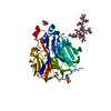
| Structure viewer | Molecule: MolmilJmol/JSmol |
|---|
- Downloads & links
-Download
| PDBx/mmCIF format | 1goj.cif.gz | 83.7 KB | Display | PDBx/mmCIF format |
|---|---|---|---|---|
| PDB format | pdb1goj.ent.gz | 62 KB | Display | PDB format |
| PDBx/mmJSON format | 1goj.json.gz | Tree view | PDBx/mmJSON format | |
| Others |  Other downloads Other downloads |
-Validation report
| Arichive directory | https://data.pdbj.org/pub/pdb/validation_reports/go/1gojftp://data.pdbj.org/pub/pdb/validation_reports/go/1goj | HTTPS FTP |
|---|
-Related structure data
| Related structure data |  1036C  2kinS S: Starting model for refinement C: citing same article ( |
|---|---|
| Similar structure data |


-Links
 PDBj
PDBj


- Assembly
| Deposited unit | 
| ||||||||
|---|---|---|---|---|---|---|---|---|---|
| 1 |
| ||||||||
| Unit cell |
|
-Components
| #1: Protein | / KINESIN Mass: 38792.836 Da / Num. of mol.: 1 / Fragment: MOTOR DOMAIN, RESIDUES 1-355 Source method: isolated from a genetically manipulated source Source: (gene. exp.) NEUROSPORA CRASSA (fungus) / Plasmid: PET21A VARIANT / Production host:  ESCHERICHIA COLI (E. coli) / Strain (production host): BL21(DE3) / References: UniProt: P48467, EC: 3.6.4.4 ESCHERICHIA COLI (E. coli) / Strain (production host): BL21(DE3) / References: UniProt: P48467, EC: 3.6.4.4 |
|---|---|
| #2: Chemical | ChemComp-ADP / Adenosine diphosphate  Mass: 427.201 Da / Num. of mol.: 1 / Source method: obtained synthetically / Formula: C10H15N5O10P2 / Comment: ADP, energy-carrying molecule*YM Mass: 427.201 Da / Num. of mol.: 1 / Source method: obtained synthetically / Formula: C10H15N5O10P2 / Comment: ADP, energy-carrying molecule*YM |
| #3: Chemical | ChemComp-MG /   Mass: 24.305 Da / Num. of mol.: 1 / Source method: obtained synthetically / Formula: Mg Mass: 24.305 Da / Num. of mol.: 1 / Source method: obtained synthetically / Formula: Mg |
| #4: Water | ChemComp-HOH / Water Mass: 18.015 Da / Num. of mol.: 114 / Source method: isolated from a natural source / Formula: H2O Mass: 18.015 Da / Num. of mol.: 114 / Source method: isolated from a natural source / Formula: H2O |
-Experimental details
-Experiment
| Experiment | Method: X-RAY DIFFRACTION / Number of used crystals: 1 |
|---|
- Sample preparation
| Crystal | Density Matthews: 2.07 Å3/Da / Density % sol: 40.6 % | ||||||||||||||||||||||||||||||
|---|---|---|---|---|---|---|---|---|---|---|---|---|---|---|---|---|---|---|---|---|---|---|---|---|---|---|---|---|---|---|---|
| Crystal grow | pH: 7 Details: 100 MM HEPES PH 6.5-7.5, 17.5% PEGMME2000, 3% GLYCEROL, PROTEIN CONCENTRATION = 7.5 - 15 MG/ML | ||||||||||||||||||||||||||||||
| Crystal grow | *PLUS Temperature: 19 ℃ / Method: vapor diffusion, sitting drop | ||||||||||||||||||||||||||||||
| Components of the solutions | *PLUS
|
-Data collection
| Diffraction | Mean temperature: 93 K |
|---|---|
| Diffraction source | Source: SYNCHROTRON / Site: MPG/DESY, HAMBURG / Beamline: BW6 / Wavelength: 1.05 |
| Detector | Type: MARRESEARCH / Detector: CCD / Date: Jun 15, 2000 / Details: MIRROR |
| Radiation | Monochromator: SILICON 111 / Protocol: SINGLE WAVELENGTH / Monochromatic (M) / Laue (L): M / Scattering type: x-ray |
| Radiation wavelength | Wavelength: 1.05 Å / Relative weight: 1 |
| Reflection | Resolution: 2.3→30 Å / Num. obs: 14829 / % possible obs: 99.8 % / Redundancy: 8 % / Biso Wilson estimate: 17.4 Å2 / Rmerge(I) obs: 0.099 / Net I/σ(I): 15.8 |
| Reflection shell | Resolution: 2.3→2.4 Å / Redundancy: 9.4 % / Rmerge(I) obs: 0.319 / Mean I/σ(I) obs: 6.3 / % possible all: 99.8 |
| Reflection | *PLUS Lowest resolution: 30 Å / Redundancy: 8 % / Num. measured all: 138093 |
| Reflection shell | *PLUS % possible obs: 100 % |
- Processing
| Software |
| ||||||||||||||||||||||||||||||||||||||||||||||||||||||||||||||||||||||||||||||||
|---|---|---|---|---|---|---|---|---|---|---|---|---|---|---|---|---|---|---|---|---|---|---|---|---|---|---|---|---|---|---|---|---|---|---|---|---|---|---|---|---|---|---|---|---|---|---|---|---|---|---|---|---|---|---|---|---|---|---|---|---|---|---|---|---|---|---|---|---|---|---|---|---|---|---|---|---|---|---|---|---|---|
| Refinement | Method to determine structure: MOLECULAR REPLACEMENT Starting model: 2KIN Resolution: 2.3→30 Å / Rfactor Rfree error: 0.009 / Data cutoff high absF: 3158928.54 / Isotropic thermal model: RESTRAINED / Cross valid method: THROUGHOUT / σ(F): 0
| ||||||||||||||||||||||||||||||||||||||||||||||||||||||||||||||||||||||||||||||||
| Solvent computation | Solvent model: FLAT MODEL / Bsol: 34 Å2 / ksol: 0.380799 e/Å3 | ||||||||||||||||||||||||||||||||||||||||||||||||||||||||||||||||||||||||||||||||
| Displacement parameters | Biso mean: 30.4 Å2
| ||||||||||||||||||||||||||||||||||||||||||||||||||||||||||||||||||||||||||||||||
| Refine analyze |
| ||||||||||||||||||||||||||||||||||||||||||||||||||||||||||||||||||||||||||||||||
| Refinement step | Cycle: LAST / Resolution: 2.3→30 Å
| ||||||||||||||||||||||||||||||||||||||||||||||||||||||||||||||||||||||||||||||||
| Refine LS restraints |
| ||||||||||||||||||||||||||||||||||||||||||||||||||||||||||||||||||||||||||||||||
| LS refinement shell | Resolution: 2.3→2.44 Å / Rfactor Rfree error: 0.024 / Total num. of bins used: 6
| ||||||||||||||||||||||||||||||||||||||||||||||||||||||||||||||||||||||||||||||||
| Xplor file |
| ||||||||||||||||||||||||||||||||||||||||||||||||||||||||||||||||||||||||||||||||
| Refinement | *PLUS Lowest resolution: 30 Å / % reflection Rfree: 10 % / Rfactor obs: 0.219 / Rfactor Rfree: 0.256 / Rfactor Rwork: 0.219 | ||||||||||||||||||||||||||||||||||||||||||||||||||||||||||||||||||||||||||||||||
| Solvent computation | *PLUS | ||||||||||||||||||||||||||||||||||||||||||||||||||||||||||||||||||||||||||||||||
| Displacement parameters | *PLUS | ||||||||||||||||||||||||||||||||||||||||||||||||||||||||||||||||||||||||||||||||
| Refine LS restraints | *PLUS
| ||||||||||||||||||||||||||||||||||||||||||||||||||||||||||||||||||||||||||||||||
| LS refinement shell | *PLUS Total num. of bins used: 6 |
