 Movie
Movie Controller


+ Open data

- Basic information
| Entry | Database: PDB / ID: 1gl2 | |||||||||
|---|---|---|---|---|---|---|---|---|---|---|
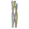
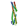
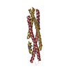
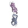
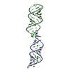
| Title | Crystal structure of an endosomal SNARE core complex | |||||||||
 Components Components |
| |||||||||
 Keywords Keywords |  MEMBRANE PROTEIN / MEMBRANE FUSION PROTEIN COMPLEX / COILED COIL / TRANSMEMBRANE / TRANSPORT MEMBRANE PROTEIN / MEMBRANE FUSION PROTEIN COMPLEX / COILED COIL / TRANSMEMBRANE / TRANSPORT | |||||||||
| Function / homology |  Function and homology information Function and homology informationzymogen granule exocytosis / positive regulation of pancreatic amylase secretion / organelle localization / tertiary granule / mucin granule / negative regulation of secretion by cell / positive regulation of receptor localization to synapse / positive regulation of histamine secretion by mast cell / : / zymogen granule ...zymogen granule exocytosis / positive regulation of pancreatic amylase secretion / organelle localization / tertiary granule / mucin granule / negative regulation of secretion by cell / positive regulation of receptor localization to synapse / positive regulation of histamine secretion by mast cell / : / zymogen granule / vesicle fusion with Golgi apparatus / organelle assembly / Lysosome Vesicle Biogenesis / Golgi Associated Vesicle Biogenesis / Golgi to vacuole transport / mucus secretion / vesicle fusion / vesicle docking / Platelet degranulation / chloride channel inhibitor activity / SNARE complex / SNAP receptor activity / intra-Golgi vesicle-mediated transport / Cargo recognition for clathrin-mediated endocytosis / Clathrin-mediated endocytosis / early endosome to late endosome transport / retrograde transport, endosome to Golgi / SNARE complex assembly / syntaxin binding / Neutrophil degranulation / azurophil granule / azurophil granule membrane / autophagosome membrane docking / endosome to lysosome transport / autophagosome maturation / regulation of endocytosis / endocytic vesicle / immunological synapse / endomembrane system / regulation of protein localization to plasma membrane / phagocytic vesicle / vesicle-mediated transport / SNARE binding / secretory granule membrane / secretory granule / macroautophagy / intracellular protein transport / trans-Golgi network / ER to Golgi transport vesicle membrane / recycling endosome / positive regulation of T cell mediated cytotoxicity / cellular response to type II interferon / recycling endosome membrane / synaptic vesicle / late endosome / protein transport / late endosome membrane / midbody / early endosome membrane / basolateral plasma membrane / protein-containing complex assembly / defense response to virus / vesicle / membrane fusion / lysosome / membrane => GO:0016020 / early endosome / endosome / symbiont entry into host cell / lysosomal membrane / intracellular membrane-bounded organelle / neuronal cell body / ubiquitin protein ligase binding / protein-containing complex binding / endoplasmic reticulum membrane / perinuclear region of cytoplasm / Golgi apparatus / cell surface / membrane / plasma membrane / cytosol / cytoplasmSimilarity search - Function | |||||||||
| Biological species |  RATTUS NORVEGICUS (Norway rat)MUS MUSCULUS (house mouse) RATTUS NORVEGICUS (Norway rat)MUS MUSCULUS (house mouse) | |||||||||
| Method | X-RAY DIFFRACTION / MOLECULAR REPLACEMENT / Resolution: 1.9 Å | |||||||||
 Authors Authors | Antonin, W. / Becker, S. / Jahn, R. / Schneider, T.R. | |||||||||
 Citation Citation | Journal: Nat.Struct.Biol. / Year: 2001 Title: Crystal Structure of the Endosomal Snare Complex Reveals Common Structural Principles of All Snares. Authors: Antonin, W. / Fasshauer, D. / Becker, S. / Jahn, R. / Schneider, T.R. | |||||||||
| History |
|
- Structure visualization
| Structure viewer | Molecule: MolmilJmol/JSmol |
|---|
- Downloads & links
-Download
| PDBx/mmCIF format | 1gl2.cif.gz | 61.1 KB | Display | PDBx/mmCIF format |
|---|---|---|---|---|
| PDB format | pdb1gl2.ent.gz | 44.8 KB | Display | PDB format |
| PDBx/mmJSON format | 1gl2.json.gz | Tree view | PDBx/mmJSON format | |
| Others |  Other downloads Other downloads |
-Validation report
| Arichive directory | https://data.pdbj.org/pub/pdb/validation_reports/gl/1gl2ftp://data.pdbj.org/pub/pdb/validation_reports/gl/1gl2 | HTTPS FTP |
|---|
-Related structure data
| Related structure data | 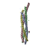 1sfcS S: Starting model for refinement |
|---|---|
| Similar structure data |


-Links
 PDBj
PDBj


- Assembly
| Deposited unit | 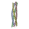
| ||||||||
|---|---|---|---|---|---|---|---|---|---|
| 1 |
| ||||||||
| Unit cell |
|
-Components
| #1: Protein | Vesicle-associated membrane protein 8 Mass: 7361.149 Da / Num. of mol.: 1 / Fragment: CORE FRAGMENT, RESIDUES 6-66 Source method: isolated from a genetically manipulated source Source: (gene. exp.) RATTUS NORVEGICUS (Norway rat) / Production host:  ESCHERICHIA COLI (E. coli) / References: UniProt: Q9WUF4 ESCHERICHIA COLI (E. coli) / References: UniProt: Q9WUF4 |
|---|---|
| #2: Protein | Mass: 7325.043 Da / Num. of mol.: 1 / Fragment: CORE FRAGMENT, RESIDUES 169-229 Source method: isolated from a genetically manipulated source Source: (gene. exp.) MUS MUSCULUS (house mouse) / Production host: ESCHERICHIA COLI (E. coli) / References: UniProt: O70439 |
| #3: Protein | Mass: 7551.417 Da / Num. of mol.: 1 / Fragment: CORE FRAGMENT, RESIDUES 140-200 Source method: isolated from a genetically manipulated source Source: (gene. exp.) MUS MUSCULUS (house mouse) / Production host: ESCHERICHIA COLI (E. coli) / References: UniProt: O88384 |
| #4: Protein | Mass: 7346.017 Da / Num. of mol.: 1 / Fragment: CORE FRAGMENT, RESIDUES 149-209 Source method: isolated from a genetically manipulated source Source: (gene. exp.) RATTUS NORVEGICUS (Norway rat) / Production host: ESCHERICHIA COLI (E. coli) / References: UniProt: Q9Z2Q7 |
| #5: Water | ChemComp-HOH / Water Mass: 18.015 Da / Num. of mol.: 234 / Source method: isolated from a natural source / Formula: H2O Mass: 18.015 Da / Num. of mol.: 234 / Source method: isolated from a natural source / Formula: H2O |
-Experimental details
-Experiment
| Experiment | Method: X-RAY DIFFRACTION / Number of used crystals: 1 |
|---|
- Sample preparation
| Crystal | Density % sol: 30 % | ||||||||||||||||||||||||||||||||||||
|---|---|---|---|---|---|---|---|---|---|---|---|---|---|---|---|---|---|---|---|---|---|---|---|---|---|---|---|---|---|---|---|---|---|---|---|---|---|
| Crystal grow | Method: vapor diffusion, hanging drop / pH: 5.2 Details: PROTEIN SOLUTION: 20 MG/ML OF QUATERNARY COMPLEX IN 20MM TRIS-HCL AT PH 7.4 WELL SOLUTION: 0.1M NA-ACETATE AT PH 5.2, 2.5 M NA-FORMIAT, 15% GLYCEROL HANGING DROPS WITH 2MUL + 2 MUL | ||||||||||||||||||||||||||||||||||||
| Crystal grow | *PLUS Temperature: 20 ℃ / pH: 7.4 / Method: vapor diffusion, hanging drop | ||||||||||||||||||||||||||||||||||||
| Components of the solutions | *PLUS
|
-Data collection
| Diffraction | Mean temperature: 100 K |
|---|---|
| Diffraction source | Source: ROTATING ANODE / Type: SIEMENS M18X / Wavelength: 1.5418 |
| Detector | Type: MAR scanner 345 mm plate / Detector: IMAGE PLATE / Date: May 15, 2001 / Details: COSMIC MIRRORS CMF12-38CU6 |
| Radiation | Monochromator: COSMIC MIRRORS CMF12-38CU6 / Protocol: SINGLE WAVELENGTH / Monochromatic (M) / Laue (L): M / Scattering type: x-ray |
| Radiation wavelength | Wavelength: 1.5418 Å / Relative weight: 1 |
| Reflection | Resolution: 1.9→15 Å / Num. obs: 71259 / % possible obs: 93.7 % / Redundancy: 5.5 % / Biso Wilson estimate: 14.3 Å2 / Rmerge(I) obs: 0.0345 / Net I/σ(I): 30.57 |
| Reflection shell | Resolution: 1.9→2 Å / Redundancy: 4.6 % / Rmerge(I) obs: 0.137 / Mean I/σ(I) obs: 11.35 / % possible all: 88.8 |
| Reflection | *PLUS Num. obs: 14969 / Num. measured all: 71259 / Rmerge(I) obs: 0.035 |
| Reflection shell | *PLUS % possible obs: 88.8 % / Redundancy: 3.6 % / Mean I/σ(I) obs: 11.4 |
- Processing
| Software |
| ||||||||||||||||||||||||||||||||||||||||||||||||||||||||||||||||||||||||||||||||
|---|---|---|---|---|---|---|---|---|---|---|---|---|---|---|---|---|---|---|---|---|---|---|---|---|---|---|---|---|---|---|---|---|---|---|---|---|---|---|---|---|---|---|---|---|---|---|---|---|---|---|---|---|---|---|---|---|---|---|---|---|---|---|---|---|---|---|---|---|---|---|---|---|---|---|---|---|---|---|---|---|---|
| Refinement | Method to determine structure: MOLECULAR REPLACEMENT Starting model: PDB ENTRY 1SFC Resolution: 1.9→14.9 Å / Rfactor Rfree error: 0.008 / Data cutoff high absF: 0 / Isotropic thermal model: RESTRAINED / Cross valid method: THROUGHOUT / σ(F): 0 / Stereochemistry target values: MLF
| ||||||||||||||||||||||||||||||||||||||||||||||||||||||||||||||||||||||||||||||||
| Solvent computation | Solvent model: FLAT MODEL / Bsol: 66.7879 Å2 / ksol: 0.418194 e/Å3 | ||||||||||||||||||||||||||||||||||||||||||||||||||||||||||||||||||||||||||||||||
| Displacement parameters | Biso mean: 22.1 Å2
| ||||||||||||||||||||||||||||||||||||||||||||||||||||||||||||||||||||||||||||||||
| Refine analyze |
| ||||||||||||||||||||||||||||||||||||||||||||||||||||||||||||||||||||||||||||||||
| Refinement step | Cycle: LAST / Resolution: 1.9→14.9 Å
| ||||||||||||||||||||||||||||||||||||||||||||||||||||||||||||||||||||||||||||||||
| Refine LS restraints |
| ||||||||||||||||||||||||||||||||||||||||||||||||||||||||||||||||||||||||||||||||
| LS refinement shell | Resolution: 1.9→2.02 Å / Rfactor Rfree error: 0.025 / Total num. of bins used: 6
| ||||||||||||||||||||||||||||||||||||||||||||||||||||||||||||||||||||||||||||||||
| Xplor file |
| ||||||||||||||||||||||||||||||||||||||||||||||||||||||||||||||||||||||||||||||||
| Software | *PLUS Name: CNS / Version: 1 / Classification: refinement | ||||||||||||||||||||||||||||||||||||||||||||||||||||||||||||||||||||||||||||||||
| Refinement | *PLUS Lowest resolution: 15 Å | ||||||||||||||||||||||||||||||||||||||||||||||||||||||||||||||||||||||||||||||||
| Solvent computation | *PLUS | ||||||||||||||||||||||||||||||||||||||||||||||||||||||||||||||||||||||||||||||||
| Displacement parameters | *PLUS | ||||||||||||||||||||||||||||||||||||||||||||||||||||||||||||||||||||||||||||||||
| Refine LS restraints | *PLUS
|
