 Movie
Movie Controller


+ Open data

- Basic information
| Entry | Database: PDB / ID: 1g9o | ||||||
|---|---|---|---|---|---|---|---|
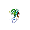
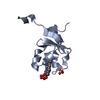
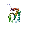
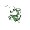
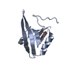
| Title | FIRST PDZ DOMAIN OF THE HUMAN NA+/H+ EXCHANGER REGULATORY FACTOR | ||||||
 Components Components | NHE-RF | ||||||
 Keywords Keywords |  SIGNALING PROTEIN / PDZ domain / NHERF / COMPLEX SIGNALING PROTEIN / PDZ domain / NHERF / COMPLEX | ||||||
| Function / homology |  Function and homology informationdopamine receptor binding / import across plasma membrane / channel activator activity / regulation of renal phosphate excretion / renal sodium ion transport / type 2 metabotropic glutamate receptor binding / glutathione transport / renal phosphate ion absorption / gamma-aminobutyric acid transmembrane transporter activity / gamma-aminobutyric acid import ...dopamine receptor binding / import across plasma membrane / channel activator activity / regulation of renal phosphate excretion / renal sodium ion transport / type 2 metabotropic glutamate receptor binding / glutathione transport / renal phosphate ion absorption / gamma-aminobutyric acid transmembrane transporter activity / gamma-aminobutyric acid import / cerebrospinal fluid circulation / negative regulation of sodium ion transport / microvillus assembly / positive regulation of monoatomic ion transmembrane transport / bile acid secretion / maintenance of epithelial cell apical/basal polarity / plasma membrane organization / stereocilium tip / cilium organization / intracellular phosphate ion homeostasis / gland morphogenesis / myosin II binding / phospholipase C-activating dopamine receptor signaling pathway / growth factor receptor binding / establishment of Golgi localization / fibroblast migration / type 3 metabotropic glutamate receptor binding / plasma membrane protein complex / establishment of epithelial cell apical/basal polarity / negative regulation of fibroblast migration / chloride channel regulator activity / negative regulation of platelet-derived growth factor receptor signaling pathway / auditory receptor cell stereocilium organization / nuclear migration / regulation of protein kinase activity / beta-2 adrenergic receptor binding / microvillus membrane / regulation of cell size / renal absorption / microvillus / transport across blood-brain barrier / negative regulation of mitotic cell cycle / negative regulation of phosphatidylinositol 3-kinase/protein kinase B signal transduction / phosphatase binding / endomembrane system / positive regulation of intrinsic apoptotic signaling pathway / protein-membrane adaptor activity / sperm midpiece / ruffle / filopodium / protein localization to plasma membrane / cell periphery / PDZ domain binding / morphogenesis of an epithelium / brush border membrane / sensory perception of sound / negative regulation of canonical Wnt signaling pathway / negative regulation of ERK1 and ERK2 cascade / beta-catenin binding / Wnt signaling pathway / adenylate cyclase-activating dopamine receptor signaling pathway / : / actin cytoskeleton / regulation of cell shape / actin cytoskeleton organization / protein-containing complex assembly / vesicle / transmembrane transporter binding / apical plasma membrane / negative regulation of cell population proliferation / signaling receptor binding / protein-containing complex binding / perinuclear region of cytoplasm / extracellular exosome / membrane / identical protein binding / cytoplasm Function and homology informationdopamine receptor binding / import across plasma membrane / channel activator activity / regulation of renal phosphate excretion / renal sodium ion transport / type 2 metabotropic glutamate receptor binding / glutathione transport / renal phosphate ion absorption / gamma-aminobutyric acid transmembrane transporter activity / gamma-aminobutyric acid import ...dopamine receptor binding / import across plasma membrane / channel activator activity / regulation of renal phosphate excretion / renal sodium ion transport / type 2 metabotropic glutamate receptor binding / glutathione transport / renal phosphate ion absorption / gamma-aminobutyric acid transmembrane transporter activity / gamma-aminobutyric acid import / cerebrospinal fluid circulation / negative regulation of sodium ion transport / microvillus assembly / positive regulation of monoatomic ion transmembrane transport / bile acid secretion / maintenance of epithelial cell apical/basal polarity / plasma membrane organization / stereocilium tip / cilium organization / intracellular phosphate ion homeostasis / gland morphogenesis / myosin II binding / phospholipase C-activating dopamine receptor signaling pathway / growth factor receptor binding / establishment of Golgi localization / fibroblast migration / type 3 metabotropic glutamate receptor binding / plasma membrane protein complex / establishment of epithelial cell apical/basal polarity / negative regulation of fibroblast migration / chloride channel regulator activity / negative regulation of platelet-derived growth factor receptor signaling pathway / auditory receptor cell stereocilium organization / nuclear migration / regulation of protein kinase activity / beta-2 adrenergic receptor binding / microvillus membrane / regulation of cell size / renal absorption / microvillus / transport across blood-brain barrier / negative regulation of mitotic cell cycle / negative regulation of phosphatidylinositol 3-kinase/protein kinase B signal transduction / phosphatase binding / endomembrane system / positive regulation of intrinsic apoptotic signaling pathway / protein-membrane adaptor activity / sperm midpiece / ruffle / filopodium / protein localization to plasma membrane / cell periphery / PDZ domain binding / morphogenesis of an epithelium / brush border membrane / sensory perception of sound / negative regulation of canonical Wnt signaling pathway / negative regulation of ERK1 and ERK2 cascade / beta-catenin binding / Wnt signaling pathway / adenylate cyclase-activating dopamine receptor signaling pathway / : / actin cytoskeleton / regulation of cell shape / actin cytoskeleton organization / protein-containing complex assembly / vesicle / transmembrane transporter binding / apical plasma membrane / negative regulation of cell population proliferation / signaling receptor binding / protein-containing complex binding / perinuclear region of cytoplasm / extracellular exosome / membrane / identical protein binding / cytoplasmSimilarity search - Function | ||||||
| Biological species |  Homo sapiens (human) Homo sapiens (human) | ||||||
| Method | X-RAY DIFFRACTION / SYNCHROTRON / MAD / Resolution: 1.5 Å | ||||||
 Authors Authors | Karthikeyan, S. / Leung, T. / Birrane, G. / Webster, G. / Ladias, J.A.A. | ||||||
 Citation Citation | Journal: J.Mol.Biol. / Year: 2001 Title: Crystal structure of the PDZ1 domain of human Na(+)/H(+) exchanger regulatory factor provides insights into the mechanism of carboxyl-terminal leucine recognition by class I PDZ domains. Authors: Karthikeyan, S. / Leung, T. / Birrane, G. / Webster, G. / Ladias, J.A. #1: Journal: J.Biol.Chem. / Year: 1998Title: NHE-RF, a regulatory cofactor for Na(+)-H+ exchange, is a common interactor for merlin and ERM (MERM) proteins; Authors: MURTHY, A. / GONZALEZ-AGOSTI, C. / CORDERO, E. / PINNEY, D. / CANDIA, C. / SOLOMON, F. / GUSELLA, J. / RAMESH, V. #2: Journal: Nat.Struct.Biol. / Year: 1998Title: Crystal structure of the hCASK PDZ domain reveals the structural basis of class II PDZ domain target recognition Authors: Daniels, D.L. / Cohen, A.R. / Anderson, J.M. / Brunger, A.T. #3: Journal: Cell(Cambridge,Mass.) / Year: 1996Title: Crystal structures of a complexed and peptide-free membrane protein-binding domain:Molecular basis of peptide recognition by PDZ Authors: Doyle, D.A. / Lee, A. / Lewis, J. / Kim, E. / Sheng, M. / MacKinnon, R. #4: Journal: Nature / Year: 1996Title: Crystal structure of a PDZ domain Authors: Morais Cabral, J.H. / Petosa, C. / Sutcliffe, M.J. / Raza, S. / Byron, O. / Poy, F. / Marfatia, S.M. / Chishti, A.H. / Liddington, R.C. | ||||||
| History |
|
- Structure visualization
| Structure viewer | Molecule: MolmilJmol/JSmol |
|---|
- Downloads & links
-Download
| PDBx/mmCIF format | 1g9o.cif.gz | 32.8 KB | Display | PDBx/mmCIF format |
|---|---|---|---|---|
| PDB format | pdb1g9o.ent.gz | 21.8 KB | Display | PDB format |
| PDBx/mmJSON format | 1g9o.json.gz | Tree view | PDBx/mmJSON format | |
| Others |  Other downloads Other downloads |
-Validation report
| Arichive directory | https://data.pdbj.org/pub/pdb/validation_reports/g9/1g9oftp://data.pdbj.org/pub/pdb/validation_reports/g9/1g9o | HTTPS FTP |
|---|
-Related structure data
| Similar structure data |
|---|

-Links
 PDBj
PDBj- Assembly
| Deposited unit | 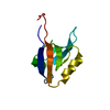
| ||||||||
|---|---|---|---|---|---|---|---|---|---|
| 1 |
| ||||||||
| Unit cell |
|
-Components
| #1: Protein | Mass: 10043.504 Da / Num. of mol.: 1 / Fragment: PDZ1 DOMAIN Source method: isolated from a genetically manipulated source Source: (gene. exp.) Homo sapiens (human) / Gene: NHERF / Plasmid: PGEX-2T / Species (production host): Escherichia coli / Production host:  Escherichia coli BL21(DE3) (bacteria) / Strain (production host): BL21(DE3) / References: UniProt: O14745 Escherichia coli BL21(DE3) (bacteria) / Strain (production host): BL21(DE3) / References: UniProt: O14745 |
|---|---|
| #2: Water | ChemComp-HOH / Water Mass: 18.015 Da / Num. of mol.: 144 / Source method: isolated from a natural source / Formula: H2O Mass: 18.015 Da / Num. of mol.: 144 / Source method: isolated from a natural source / Formula: H2O |
-Experimental details
-Experiment
| Experiment | Method: X-RAY DIFFRACTION / Number of used crystals: 1 |
|---|
- Sample preparation
| Crystal | Density Matthews: 2.3 Å3/Da / Density % sol: 48 % | ||||||||||||||||||||||||||||||||||||
|---|---|---|---|---|---|---|---|---|---|---|---|---|---|---|---|---|---|---|---|---|---|---|---|---|---|---|---|---|---|---|---|---|---|---|---|---|---|
| Crystal grow | Temperature: 291 K / Method: vapor diffusion, sitting drop / pH: 5.9 Details: potassium sodium tartrate, sodium citrate, ammonium sulfate, manganese chloride, pH 5.9, VAPOR DIFFUSION, SITTING DROP, temperature 291K | ||||||||||||||||||||||||||||||||||||
| Crystal grow | *PLUS Temperature: 20 ℃ | ||||||||||||||||||||||||||||||||||||
| Components of the solutions | *PLUS
|
-Data collection
| Diffraction | Mean temperature: 100 K |
|---|---|
| Diffraction source | Source: SYNCHROTRON / Site: CHESS  / Beamline: F2 / Wavelength: 0.98401 Å / Beamline: F2 / Wavelength: 0.98401 Å |
| Detector | Type: ADSC QUANTUM 4 / Detector: CCD / Date: May 20, 2000 / Details: DUAL SLITS |
| Radiation | Monochromator: SAGITALLY FOCUSED Si(111) / Protocol: SINGLE WAVELENGTH / Monochromatic (M) / Laue (L): M / Scattering type: x-ray |
| Radiation wavelength | Wavelength: 0.98401 Å / Relative weight: 1 |
| Reflection | Resolution: 1.5→36 Å / Num. all: 14916 / Num. obs: 14916 / % possible obs: 99.7 % / Observed criterion σ(F): -3 / Observed criterion σ(I): -3 / Redundancy: 7.5 % / Biso Wilson estimate: 16.5 Å2 / Rmerge(I) obs: 0.039 / Rsym value: 3.9 / Net I/σ(I): 35 |
| Reflection shell | Resolution: 1.5→1.55 Å / Redundancy: 4.6 % / Rmerge(I) obs: 0.152 / Mean I/σ(I) obs: 10.3 / Num. unique all: 1449 / Rsym value: 15.2 / % possible all: 98.9 |
| Reflection | *PLUS Num. measured all: 246628 |
| Reflection shell | *PLUS % possible obs: 98.9 % |
- Processing
| Software |
| ||||||||||||||||||||||||||||||||||||||||
|---|---|---|---|---|---|---|---|---|---|---|---|---|---|---|---|---|---|---|---|---|---|---|---|---|---|---|---|---|---|---|---|---|---|---|---|---|---|---|---|---|---|
| Refinement | Method to determine structure: MAD / Resolution: 1.5→24.6 Å / Rfactor Rfree error: 0.005 / Isotropic thermal model: isotropic / Cross valid method: THROUGHOUT / σ(F): 0 / σ(I): 0 / Stereochemistry target values: Engh & Huber
| ||||||||||||||||||||||||||||||||||||||||
| Solvent computation | Solvent model: FLAT MODEL / Bsol: 63.19 Å2 / ksol: 0.4078 e/Å3 | ||||||||||||||||||||||||||||||||||||||||
| Displacement parameters | Biso mean: 16.7 Å2
| ||||||||||||||||||||||||||||||||||||||||
| Refine analyze |
| ||||||||||||||||||||||||||||||||||||||||
| Refinement step | Cycle: LAST / Resolution: 1.5→24.6 Å
| ||||||||||||||||||||||||||||||||||||||||
| Refine LS restraints |
| ||||||||||||||||||||||||||||||||||||||||
| LS refinement shell | Resolution: 1.5→1.59 Å / Rfactor Rfree error: 0.015 / Total num. of bins used: 6
| ||||||||||||||||||||||||||||||||||||||||
| Xplor file |
| ||||||||||||||||||||||||||||||||||||||||
| Software | *PLUS Name: CNS / Version: 0.9 / Classification: refinement | ||||||||||||||||||||||||||||||||||||||||
| Refinement | *PLUS σ(F): 0 / % reflection Rfree: 10 % / Rfactor obs: 0.181 | ||||||||||||||||||||||||||||||||||||||||
| Solvent computation | *PLUS | ||||||||||||||||||||||||||||||||||||||||
| Displacement parameters | *PLUS Biso mean: 16.7 Å2 | ||||||||||||||||||||||||||||||||||||||||
| Refine LS restraints | *PLUS
| ||||||||||||||||||||||||||||||||||||||||
| LS refinement shell | *PLUS Rfactor Rfree: 0.232 / % reflection Rfree: 9.9 % / Rfactor Rwork: 0.189 |
