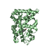
 Movie
Movie Controller


+ Open data

- Basic information
| Entry | Database: PDB / ID: 1g8a | ||||||
|---|---|---|---|---|---|---|---|
| Title | PYROCOCCUS HORIKOSHII FIBRILLARIN PRE-RRNA PROCESSING PROTEIN | ||||||
 Components Components | FIBRILLARIN-LIKE PRE-RRNA PROCESSING PROTEIN | ||||||
 Keywords Keywords |  RNA BINDING PROTEIN / rRNA binding / RNA binding / Structural Genomics / BSGC structure funded by NIH / Protein Structure Initiative / PSI / Berkeley Structural Genomics Center RNA BINDING PROTEIN / rRNA binding / RNA binding / Structural Genomics / BSGC structure funded by NIH / Protein Structure Initiative / PSI / Berkeley Structural Genomics Center | ||||||
| Function / homology |  Function and homology information Function and homology informationtRNA processing / Transferases; Transferring one-carbon groups; Methyltransferases / methyltransferase activity / rRNA processing / methylation / RNA bindingSimilarity search - Function | ||||||
| Biological species |   Pyrococcus horikoshii (archaea) Pyrococcus horikoshii (archaea) | ||||||
| Method | X-RAY DIFFRACTION / SYNCHROTRON / MOLECULAR REPLACEMENT / Resolution: 1.4 Å | ||||||
 Authors Authors | Boisvert, D.C. / Kim, S.H. / Berkeley Structural Genomics Center (BSGC) | ||||||
 Citation Citation | Journal: To be Published Title: A Structural Approach to Gene Function and Structure Quality for Pyrococcus horikoshii Fibrillarin Authors: Boisvert, D.C. / Kim, S.H. | ||||||
| History |
|
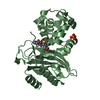
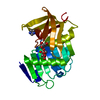
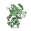
- Structure visualization
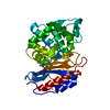
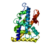
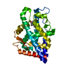
| Structure viewer | Molecule: MolmilJmol/JSmol |
|---|
- Downloads & links
-Download
| PDBx/mmCIF format | 1g8a.cif.gz | 66.3 KB | Display | PDBx/mmCIF format |
|---|---|---|---|---|
| PDB format | pdb1g8a.ent.gz | 48.1 KB | Display | PDB format |
| PDBx/mmJSON format | 1g8a.json.gz | Tree view | PDBx/mmJSON format | |
| Others |  Other downloads Other downloads |
-Validation report
| Arichive directory | https://data.pdbj.org/pub/pdb/validation_reports/g8/1g8aftp://data.pdbj.org/pub/pdb/validation_reports/g8/1g8a | HTTPS FTP |
|---|
-Related structure data
| Related structure data |  1fibS S: Starting model for refinement |
|---|---|
| Similar structure data | |
| Other databases |


-Links
 PDBj
PDBj- Assembly
| Deposited unit | 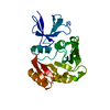
| ||||||||
|---|---|---|---|---|---|---|---|---|---|
| 1 |
| ||||||||
| Unit cell |
|
-Components
| #1: Protein | Mass: 25864.746 Da / Num. of mol.: 1 Source method: isolated from a genetically manipulated source Source: (gene. exp.) Pyrococcus horikoshii (archaea) / Gene: 0048 / Plasmid: PET21A / Species (production host): Escherichia coli / Production host:  Escherichia coli BL21(DE3) (bacteria) / Strain (production host): BL21(DE3) / References: UniProt: O57811 Escherichia coli BL21(DE3) (bacteria) / Strain (production host): BL21(DE3) / References: UniProt: O57811 |
|---|---|
| #2: Water | ChemComp-HOH / Water Mass: 18.015 Da / Num. of mol.: 407 / Source method: isolated from a natural source / Formula: H2O Mass: 18.015 Da / Num. of mol.: 407 / Source method: isolated from a natural source / Formula: H2O |
-Experimental details
-Experiment
| Experiment | Method: X-RAY DIFFRACTION / Number of used crystals: 1 |
|---|
- Sample preparation
| Crystal | Density Matthews: 1.97 Å3/Da / Density % sol: 37.69 % |
|---|---|
| Crystal grow | Temperature: 277 K / Method: cooling / pH: 7.5 Details: 50 mM NaCl, 50 mM Tris, 85 mg/ml fibrillarin, pH 7.5, Cooling, temperature 4K |
-Data collection
| Diffraction | Mean temperature: 180 K |
|---|---|
| Diffraction source | Source: SYNCHROTRON / Site: ALS  / Beamline: 5.0.2 / Wavelength: 0.95 Å / Beamline: 5.0.2 / Wavelength: 0.95 Å |
| Detector | Type: ADSC QUANTUM 4 / Detector: CCD / Date: Jun 4, 2000 |
| Radiation | Monochromator: Standard at ALS 5.0.2 / Protocol: SINGLE WAVELENGTH / Monochromatic (M) / Laue (L): M / Scattering type: x-ray |
| Radiation wavelength | Wavelength: 0.95 Å / Relative weight: 1 |
| Reflection | Resolution: 1.4→40 Å / Num. all: 43002 / Num. obs: 37696 / % possible obs: 94.2 % / Observed criterion σ(F): 0 / Observed criterion σ(I): 0 / Redundancy: 3 % / Biso Wilson estimate: 10.9 Å2 / Rmerge(I) obs: 0.033 / Net I/σ(I): 40 |
| Reflection shell | Resolution: 1.4→1.49 Å / Redundancy: 2 % / Rmerge(I) obs: 0.06 / % possible all: 71.2 |
- Processing
| Software |
| ||||||||||||||||||||||||||||||||||||
|---|---|---|---|---|---|---|---|---|---|---|---|---|---|---|---|---|---|---|---|---|---|---|---|---|---|---|---|---|---|---|---|---|---|---|---|---|---|
| Refinement | Method to determine structure: MOLECULAR REPLACEMENT Starting model: 1FIB Resolution: 1.4→40 Å / Rfactor Rfree error: 0.005 / Data cutoff high absF: 471722.3 / Data cutoff low absF: 0 / Isotropic thermal model: RESTRAINED / Cross valid method: THROUGHOUT / σ(F): 0 / σ(I): 0 / Stereochemistry target values: MLF
| ||||||||||||||||||||||||||||||||||||
| Solvent computation | Solvent model: FLAT MODEL / Bsol: 38.17 Å2 / ksol: 0.339 e/Å3 | ||||||||||||||||||||||||||||||||||||
| Displacement parameters | Biso mean: 13.8 Å2
| ||||||||||||||||||||||||||||||||||||
| Refine analyze |
| ||||||||||||||||||||||||||||||||||||
| Refinement step | Cycle: LAST / Resolution: 1.4→40 Å
| ||||||||||||||||||||||||||||||||||||
| Refine LS restraints |
| ||||||||||||||||||||||||||||||||||||
| LS refinement shell | Resolution: 1.4→1.49 Å / Rfactor Rfree error: 0.016 / Total num. of bins used: 6
| ||||||||||||||||||||||||||||||||||||
| Xplor file |
|
