 Movie
Movie Controller


+ Open data

- Basic information
| Entry | Database: PDB / ID: 1g6t | ||||||
|---|---|---|---|---|---|---|---|
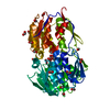
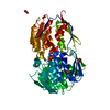
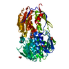
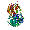
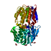
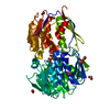
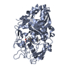
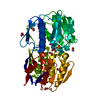
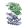
| Title | STRUCTURE OF EPSP SYNTHASE LIGANDED WITH SHIKIMATE-3-PHOSPHATE | ||||||
 Components Components | EPSP SYNTHASE | ||||||
 Keywords Keywords | TRANSFERASE / two-domain structure / inside-out alpha-beta barrel | ||||||
| Function / homology |  Function and homology information3-phosphoshikimate 1-carboxyvinyltransferase / 3-phosphoshikimate 1-carboxyvinyltransferase activity / chorismate biosynthetic process / aromatic amino acid family biosynthetic process / amino acid biosynthetic process / cytosol Function and homology information3-phosphoshikimate 1-carboxyvinyltransferase / 3-phosphoshikimate 1-carboxyvinyltransferase activity / chorismate biosynthetic process / aromatic amino acid family biosynthetic process / amino acid biosynthetic process / cytosolSimilarity search - Function | ||||||
| Biological species |  Escherichia coli (E. coli) Escherichia coli (E. coli) | ||||||
| Method | X-RAY DIFFRACTION / MOLECULAR REPLACEMENT / Resolution: 1.6 Å | ||||||
 Authors Authors | Schonbrunn, E. / Eschenburg, S. / Shuttleworth, W. / Schloss, J.V. / Amrhein, N. / Evans, J.N.S. / Kabsch, W. | ||||||
 Citation Citation | Journal: Proc.Natl.Acad.Sci.USA / Year: 2001 Title: Interaction of the herbicide glyphosate with its target enzyme 5-enolpyruvylshikimate 3-phosphate synthase in atomic detail. Authors: Schonbrunn, E. / Eschenburg, S. / Shuttleworth, W.A. / Schloss, J.V. / Amrhein, N. / Evans, J.N. / Kabsch, W. | ||||||
| History |
|
- Structure visualization
| Structure viewer | Molecule: MolmilJmol/JSmol |
|---|
- Downloads & links
-Download
| PDBx/mmCIF format | 1g6t.cif.gz | 109.1 KB | Display | PDBx/mmCIF format |
|---|---|---|---|---|
| PDB format | pdb1g6t.ent.gz | 82.4 KB | Display | PDB format |
| PDBx/mmJSON format | 1g6t.json.gz | Tree view | PDBx/mmJSON format | |
| Others |  Other downloads Other downloads |
-Validation report
| Arichive directory | https://data.pdbj.org/pub/pdb/validation_reports/g6/1g6tftp://data.pdbj.org/pub/pdb/validation_reports/g6/1g6t | HTTPS FTP |
|---|
-Related structure data


-Links
 PDBj
PDBj
- Assembly
| Deposited unit | 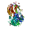
| ||||||||
|---|---|---|---|---|---|---|---|---|---|
| 1 |
| ||||||||
| Unit cell |
|
-Components
| #1: Protein | / 3-PHOSPHOSHIKIMATE 1-CARBOXYVINYLTRANSFERASE Mass: 46141.613 Da / Num. of mol.: 1 Source method: isolated from a genetically manipulated source Source: (gene. exp.) Escherichia coli (E. coli) / Gene: AROA / Production host: Escherichia coli (E. coli)References: UniProt: P0A6D3, 3-phosphoshikimate 1-carboxyvinyltransferase | ||
|---|---|---|---|
| #2: Chemical | ChemComp-PO4 / Phosphate  Mass: 94.971 Da / Num. of mol.: 1 / Source method: obtained synthetically / Formula: PO4 Mass: 94.971 Da / Num. of mol.: 1 / Source method: obtained synthetically / Formula: PO4 | ||
| #3: Chemical | ChemComp-S3P /   Mass: 254.131 Da / Num. of mol.: 1 / Source method: obtained synthetically / Formula: C7H11O8P Mass: 254.131 Da / Num. of mol.: 1 / Source method: obtained synthetically / Formula: C7H11O8P | ||
| #4: Chemical | ChemComp-FMT / Formic acid  Mass: 46.025 Da / Num. of mol.: 13 / Source method: obtained synthetically / Formula: CH2O2 Mass: 46.025 Da / Num. of mol.: 13 / Source method: obtained synthetically / Formula: CH2O2#5: Water | ChemComp-HOH / | Water Mass: 18.015 Da / Num. of mol.: 555 / Source method: isolated from a natural source / Formula: H2O Mass: 18.015 Da / Num. of mol.: 555 / Source method: isolated from a natural source / Formula: H2O |
-Experimental details
-Experiment
| Experiment | Method: X-RAY DIFFRACTION / Number of used crystals: 1 |
|---|
- Sample preparation
| Crystal | Density Matthews: 2.34 Å3/Da / Density % sol: 47.42 % | ||||||||||||||||||||||||
|---|---|---|---|---|---|---|---|---|---|---|---|---|---|---|---|---|---|---|---|---|---|---|---|---|---|
| Crystal grow | Temperature: 293 K / Method: vapor diffusion / pH: 7 / Details: pH 7, VAPOR DIFFUSION, temperature 293K | ||||||||||||||||||||||||
| Crystal grow | *PLUS Temperature: 20 ℃ / Method: vapor diffusion, hanging drop | ||||||||||||||||||||||||
| Components of the solutions | *PLUS
|
-Data collection
| Diffraction | Mean temperature: 120 K |
|---|---|
| Diffraction source | Source: ROTATING ANODE / Type: RIGAKU RU300 / Wavelength: 1.54 Å |
| Detector | Type: RIGAKU RAXIS IV / Detector: IMAGE PLATE / Date: Mar 29, 2000 / Details: osmic mirrors |
| Radiation | Monochromator: osmic mirrors / Protocol: SINGLE WAVELENGTH / Monochromatic (M) / Laue (L): M / Scattering type: x-ray |
| Radiation wavelength | Wavelength: 1.54 Å / Relative weight: 1 |
| Reflection | Resolution: 1.6→20 Å / Num. all: 57076 / Num. obs: 57076 / % possible obs: 98.6 % / Observed criterion σ(F): 0 / Observed criterion σ(I): -3 / Redundancy: 3.3 % / Biso Wilson estimate: 14.8 Å2 / Rmerge(I) obs: 0.049 / Net I/σ(I): 15.1 |
| Reflection shell | Resolution: 1.6→1.65 Å / Redundancy: 2.5 % / Rmerge(I) obs: 0.18 / Mean I/σ(I) obs: 4.7 / % possible all: 95.7 |
| Reflection | *PLUS Highest resolution: 1.6 Å / Lowest resolution: 20 Å / Num. measured all: 182207 |
| Reflection shell | *PLUS Highest resolution: 1.6 Å / % possible obs: 95.7 % / Num. unique obs: 4837 / Num. measured obs: 12801 / Rmerge(I) obs: 0.181 |
- Processing
| Software |
| ||||||||||||||||||||||||||||||||||||||||||||||||||||||||||||
|---|---|---|---|---|---|---|---|---|---|---|---|---|---|---|---|---|---|---|---|---|---|---|---|---|---|---|---|---|---|---|---|---|---|---|---|---|---|---|---|---|---|---|---|---|---|---|---|---|---|---|---|---|---|---|---|---|---|---|---|---|---|
| Refinement | Method to determine structure: MOLECULAR REPLACEMENT / Resolution: 1.6→19.94 Å / Rfactor Rfree error: 0.004 / Data cutoff high absF: 2914701.6 / Data cutoff low absF: 0 / Isotropic thermal model: RESTRAINED / Cross valid method: THROUGHOUT / σ(I): -3 / Stereochemistry target values: Engh & Huber
| ||||||||||||||||||||||||||||||||||||||||||||||||||||||||||||
| Solvent computation | Solvent model: FLAT MODEL / Bsol: 49.83 Å2 / ksol: 0.403 e/Å3 | ||||||||||||||||||||||||||||||||||||||||||||||||||||||||||||
| Displacement parameters | Biso mean: 14.3 Å2
| ||||||||||||||||||||||||||||||||||||||||||||||||||||||||||||
| Refine analyze |
| ||||||||||||||||||||||||||||||||||||||||||||||||||||||||||||
| Refinement step | Cycle: LAST / Resolution: 1.6→19.94 Å
| ||||||||||||||||||||||||||||||||||||||||||||||||||||||||||||
| Refine LS restraints |
| ||||||||||||||||||||||||||||||||||||||||||||||||||||||||||||
| LS refinement shell | Resolution: 1.6→1.7 Å / Rfactor Rfree error: 0.013 / Total num. of bins used: 6
| ||||||||||||||||||||||||||||||||||||||||||||||||||||||||||||
| Xplor file |
| ||||||||||||||||||||||||||||||||||||||||||||||||||||||||||||
| Software | *PLUS Name: CNS / Version: 1 / Classification: refinement | ||||||||||||||||||||||||||||||||||||||||||||||||||||||||||||
| Refinement | *PLUS % reflection Rfree: 3 % | ||||||||||||||||||||||||||||||||||||||||||||||||||||||||||||
| Solvent computation | *PLUS | ||||||||||||||||||||||||||||||||||||||||||||||||||||||||||||
| Displacement parameters | *PLUS Biso mean: 14.3 Å2 | ||||||||||||||||||||||||||||||||||||||||||||||||||||||||||||
| Refine LS restraints | *PLUS
| ||||||||||||||||||||||||||||||||||||||||||||||||||||||||||||
| LS refinement shell | *PLUS Rfactor Rfree: 0.222 / % reflection Rfree: 3 % / Rfactor Rwork: 0.208 |
