 Movie
Movie Controller

[English] 日本語
 Yorodumi
- PDB-1fx2: STRUCTURAL ANALYSIS OF ADENYLATE CYCLASES FROM TRYPANOSOMA BRUCEI... -
+ Open data

- Basic information
| Entry | Database: PDB / ID: 1fx2 | ||||||
|---|---|---|---|---|---|---|---|
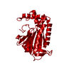
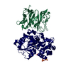
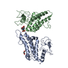
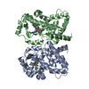
| Title | STRUCTURAL ANALYSIS OF ADENYLATE CYCLASES FROM TRYPANOSOMA BRUCEI IN THEIR MONOMERIC STATE | ||||||
 Components Components | RECEPTOR-TYPE ADENYLATE CYCLASE GRESAG 4.1 | ||||||
 Keywords Keywords |  LYASE / cAMP / trypanosomes / adenylyl cyclases / monomer-dimer / catalysis LYASE / cAMP / trypanosomes / adenylyl cyclases / monomer-dimer / catalysis | ||||||
| Function / homology |  Function and homology informationadenylate cyclase / cAMP biosynthetic process / adenylate cyclase activity / membrane => GO:0016020 / intracellular signal transduction / ATP binding / metal ion binding Function and homology informationadenylate cyclase / cAMP biosynthetic process / adenylate cyclase activity / membrane => GO:0016020 / intracellular signal transduction / ATP binding / metal ion bindingSimilarity search - Function | ||||||
| Biological species |  Trypanosoma brucei (eukaryote) Trypanosoma brucei (eukaryote) | ||||||
| Method | X-RAY DIFFRACTION / SYNCHROTRON / Resolution: 1.46 Å | ||||||
 Authors Authors | Bieger, B. / Essen, L.-O. | ||||||
 Citation Citation | Journal: EMBO J. / Year: 2001 Title: Structural analysis of adenylate cyclases from Trypanosoma brucei in their monomeric state. Authors: Bieger, B. / Essen, L.O. | ||||||
| History |
|
- Structure visualization
| Structure viewer | Molecule: MolmilJmol/JSmol |
|---|
- Downloads & links
-Download
| PDBx/mmCIF format | 1fx2.cif.gz | 67.7 KB | Display | PDBx/mmCIF format |
|---|---|---|---|---|
| PDB format | pdb1fx2.ent.gz | 49.4 KB | Display | PDB format |
| PDBx/mmJSON format | 1fx2.json.gz | Tree view | PDBx/mmJSON format | |
| Others |  Other downloads Other downloads |
-Validation report
| Arichive directory | https://data.pdbj.org/pub/pdb/validation_reports/fx/1fx2ftp://data.pdbj.org/pub/pdb/validation_reports/fx/1fx2 | HTTPS FTP |
|---|
-Related structure data


-Links
 PDBj
PDBj


- Assembly
| Deposited unit | 
| ||||||||
|---|---|---|---|---|---|---|---|---|---|
| 1 | 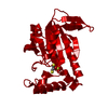
| ||||||||
| Unit cell |
| ||||||||
| Details | The biological assembly in the catalytic state is a dimer |
-Components
| #1: Protein | Mass: 26504.740 Da / Num. of mol.: 1 / Fragment: CATALYTIC DOMAIN Source method: isolated from a genetically manipulated source Source: (gene. exp.) Trypanosoma brucei (eukaryote) / Strain: STRAIN 927 / Plasmid: PET28A / Production host:  Escherichia coli (E. coli) / References: UniProt: Q99279, adenylate cyclase Escherichia coli (E. coli) / References: UniProt: Q99279, adenylate cyclase |
|---|---|
| #2: Chemical | ChemComp-SO4 / Sulfate  Mass: 96.063 Da / Num. of mol.: 1 / Source method: obtained synthetically / Formula: SO4 Mass: 96.063 Da / Num. of mol.: 1 / Source method: obtained synthetically / Formula: SO4 |
| #3: Chemical | ChemComp-DTT / Dithiothreitol  Mass: 154.251 Da / Num. of mol.: 1 / Source method: obtained synthetically / Formula: C4H10O2S2 Mass: 154.251 Da / Num. of mol.: 1 / Source method: obtained synthetically / Formula: C4H10O2S2 |
| #4: Water | ChemComp-HOH / Water Mass: 18.015 Da / Num. of mol.: 272 / Source method: isolated from a natural source / Formula: H2O Mass: 18.015 Da / Num. of mol.: 272 / Source method: isolated from a natural source / Formula: H2O |
-Experimental details
-Experiment
| Experiment | Method: X-RAY DIFFRACTION / Number of used crystals: 1 |
|---|
- Sample preparation
| Crystal | Density Matthews: 2.22 Å3/Da / Density % sol: 44.49 % | ||||||||||||||||||||||||||||||
|---|---|---|---|---|---|---|---|---|---|---|---|---|---|---|---|---|---|---|---|---|---|---|---|---|---|---|---|---|---|---|---|
| Crystal grow | Temperature: 291 K / Method: vapor diffusion / pH: 6.5 Details: 20 mM Tris/HCl, 1.8-1.9 M ammonium sulfate, pH 6.5, VAPOR DIFFUSION, temperature 18K | ||||||||||||||||||||||||||||||
| Crystal grow | *PLUS pH: 8 / Method: vapor diffusion, hanging drop | ||||||||||||||||||||||||||||||
| Components of the solutions | *PLUS
|
-Data collection
| Diffraction | Mean temperature: 100 K |
|---|---|
| Diffraction source | Source: SYNCHROTRON / Site: EMBL/DESY, HAMBURG  / Beamline: BW7B / Wavelength: 0.833 / Beamline: BW7B / Wavelength: 0.833 |
| Detector | Type: MARRESEARCH / Detector: IMAGE PLATE / Date: Dec 8, 1998 |
| Radiation | Protocol: SINGLE WAVELENGTH / Monochromatic (M) / Laue (L): M / Scattering type: x-ray |
| Radiation wavelength | Wavelength: 0.833 Å / Relative weight: 1 |
| Reflection | Resolution: 1.46→10 Å / Num. all: 498167 / Num. obs: 40911 / % possible obs: 98.7 % / Observed criterion σ(F): 0 / Observed criterion σ(I): 0 / Redundancy: 3 % / Biso Wilson estimate: 13.1 Å2 / Rmerge(I) obs: 0.082 / Net I/σ(I): 24.7 |
| Reflection shell | Resolution: 1.44→1.48 Å / Redundancy: 2.5 % / Rmerge(I) obs: 0.283 / Num. unique all: 2799 / % possible all: 98.9 |
| Reflection | *PLUS Num. measured all: 498167 |
| Reflection shell | *PLUS % possible obs: 98.9 % / Mean I/σ(I) obs: 4.3 |
- Processing
| Software |
| ||||||||||||||||||||||||||||||||||||||||
|---|---|---|---|---|---|---|---|---|---|---|---|---|---|---|---|---|---|---|---|---|---|---|---|---|---|---|---|---|---|---|---|---|---|---|---|---|---|---|---|---|---|
| Refinement | Resolution: 1.46→10 Å / Rfactor Rfree error: 0.006 / Data cutoff high absF: 1440982.24 / Data cutoff low absF: 0 / Isotropic thermal model: RESTRAINED / Cross valid method: THROUGHOUT / σ(F): 0 / σ(I): 0 / Stereochemistry target values: Engh & Huber
| ||||||||||||||||||||||||||||||||||||||||
| Solvent computation | Solvent model: FLAT MODEL / Bsol: 68.42 Å2 / ksol: 0.431 e/Å3 | ||||||||||||||||||||||||||||||||||||||||
| Displacement parameters | Biso mean: 20.1 Å2
| ||||||||||||||||||||||||||||||||||||||||
| Refine analyze |
| ||||||||||||||||||||||||||||||||||||||||
| Refinement step | Cycle: LAST / Resolution: 1.46→10 Å
| ||||||||||||||||||||||||||||||||||||||||
| Refine LS restraints |
| ||||||||||||||||||||||||||||||||||||||||
| LS refinement shell | Resolution: 1.46→1.55 Å / Rfactor Rfree error: 0.021 / Total num. of bins used: 6
| ||||||||||||||||||||||||||||||||||||||||
| Xplor file |
| ||||||||||||||||||||||||||||||||||||||||
| Software | *PLUS Name: CNS / Version: 1 / Classification: refinement | ||||||||||||||||||||||||||||||||||||||||
| Refinement | *PLUS σ(F): 0 / % reflection Rfree: 2.8 % | ||||||||||||||||||||||||||||||||||||||||
| Solvent computation | *PLUS | ||||||||||||||||||||||||||||||||||||||||
| Displacement parameters | *PLUS Biso mean: 20.1 Å2 | ||||||||||||||||||||||||||||||||||||||||
| Refine LS restraints | *PLUS
| ||||||||||||||||||||||||||||||||||||||||
| LS refinement shell | *PLUS Rfactor Rfree: 0.264 / % reflection Rfree: 2.6 % / Rfactor Rwork: 0.222 |
