 Movie
Movie Controller


+ Open data

- Basic information
| Entry | Database: PDB / ID: 1fx0 | ||||||
|---|---|---|---|---|---|---|---|
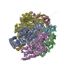
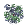
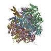
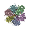
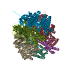
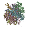
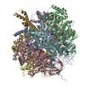
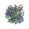
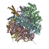
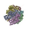
| Title | Crystal structure of the chloroplast F1-ATPase from spinach | ||||||
 Components Components |
| ||||||
 Keywords Keywords |  HYDROLASE / latent ATPase / thermal stability / potential tentoxin binding site HYDROLASE / latent ATPase / thermal stability / potential tentoxin binding site | ||||||
| Function / homology |  Function and homology information Function and homology informationmitochondrial proton-transporting ATP synthase complex / chloroplast thylakoid membrane / proton motive force-driven mitochondrial ATP synthesis / proton motive force-driven ATP synthesis / proton-transporting ATP synthase complex, catalytic core F(1) / H+-transporting two-sector ATPase / proton-transporting ATPase activity, rotational mechanism / proton-transporting ATP synthase activity, rotational mechanism / ADP binding / ATP hydrolysis activity / ATP bindingSimilarity search - Function | ||||||
| Biological species |  Spinacia oleracea (spinach) Spinacia oleracea (spinach) | ||||||
| Method | X-RAY DIFFRACTION / SYNCHROTRON / Resolution: 3.2 Å | ||||||
 Authors Authors | Groth, G. / Pohl, E. | ||||||
 Citation Citation | Journal: J.Biol.Chem. / Year: 2001 Title: The structure of the chloroplast F1-ATPase at 3.2 A resolution. Authors: Groth, G. / Pohl, E. #1: Journal: Eur.J.Biochem. / Year: 1999Title: Rapid Purification of Membrane extrinsic F1-domain of chloroplast ATP synthase in monodisperse form suitable for 3D crystallization Authors: Groth, G. / Schirwitz, K. | ||||||
| History |
|
- Structure visualization
| Structure viewer | Molecule: MolmilJmol/JSmol |
|---|
- Downloads & links
-Download
| PDBx/mmCIF format | 1fx0.cif.gz | 186.2 KB | Display | PDBx/mmCIF format |
|---|---|---|---|---|
| PDB format | pdb1fx0.ent.gz | 148.8 KB | Display | PDB format |
| PDBx/mmJSON format | 1fx0.json.gz | Tree view | PDBx/mmJSON format | |
| Others |  Other downloads Other downloads |
-Validation report
| Arichive directory | https://data.pdbj.org/pub/pdb/validation_reports/fx/1fx0ftp://data.pdbj.org/pub/pdb/validation_reports/fx/1fx0 | HTTPS FTP |
|---|
-Related structure data
| Similar structure data |
|---|
-Links
 PDBj
PDBj


- Assembly
| Deposited unit | 
| ||||||||
|---|---|---|---|---|---|---|---|---|---|
| 1 | 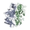
| ||||||||
| Unit cell |
| ||||||||
| Details | The biological assembly is a Hetero-Hexamer containing 3 copies of the A and 3 copies of the B subunit The asymmetric unit contains also one third of the gamma and one third of the epsilon subunit. The structures of these subunits, however, were not resolved. The heterohexamer consist of 3x(alpha), 3x(beta), 1x(gamma), 1x(epsilon). |
-Components
| #1: Protein | Mass: 55505.199 Da / Num. of mol.: 1 / Source method: isolated from a natural source / Source: (natural) Spinacia oleracea (spinach) / References: UniProt: P06450, EC: 3.6.1.34 |
|---|---|
| #2: Protein | Mass: 53921.574 Da / Num. of mol.: 1 / Source method: isolated from a natural source / Source: (natural) Spinacia oleracea (spinach) / References: UniProt: P00825, EC: 3.6.1.34 |
-Experimental details
-Experiment
| Experiment | Method: X-RAY DIFFRACTION / Number of used crystals: 1 |
|---|
- Sample preparation
| Crystal | Density Matthews: 3.69 Å3/Da / Density % sol: 66.7 % | ||||||||||||||||||||||||||||||||||||
|---|---|---|---|---|---|---|---|---|---|---|---|---|---|---|---|---|---|---|---|---|---|---|---|---|---|---|---|---|---|---|---|---|---|---|---|---|---|
| Crystal grow | Temperature: 298 K / Method: microbatch / pH: 7.5 Details: ammonium sulfate, HEPES, Na-azide, pH 7.5, Microbatch, temperature 25K | ||||||||||||||||||||||||||||||||||||
| Crystal grow | *PLUS Temperature: 20 ℃ / Method: batch method | ||||||||||||||||||||||||||||||||||||
| Components of the solutions | *PLUS
|
-Data collection
| Diffraction | Mean temperature: 100 K |
|---|---|
| Diffraction source | Source: SYNCHROTRON / Site: EMBL/DESY, HAMBURG  / Beamline: X11 / Wavelength: 0.91 / Beamline: X11 / Wavelength: 0.91 |
| Detector | Type: MARRESEARCH / Detector: CCD / Date: Feb 14, 2000 |
| Radiation | Protocol: SINGLE WAVELENGTH / Monochromatic (M) / Laue (L): M / Scattering type: x-ray |
| Radiation wavelength | Wavelength: 0.91 Å / Relative weight: 1 |
| Reflection | Resolution: 3.2→20 Å / Num. all: 23315 / Num. obs: 23050 / % possible obs: 86.2 % / Observed criterion σ(F): 0 / Observed criterion σ(I): 3 / Redundancy: 3.9 % / Biso Wilson estimate: 73.54 Å2 / Rmerge(I) obs: 0.087 / Net I/σ(I): 6.4 |
| Reflection shell | Resolution: 3.2→3.37 Å / Redundancy: 2 % / Rmerge(I) obs: 0.288 / % possible all: 76 |
| Reflection | *PLUS Lowest resolution: 20 Å / Num. measured all: 231238 |
- Processing
| Software |
| ||||||||||||||||||||
|---|---|---|---|---|---|---|---|---|---|---|---|---|---|---|---|---|---|---|---|---|---|
| Refinement | Resolution: 3.2→6 Å / σ(F): 0 / σ(I): 0 / Stereochemistry target values: Engh & Huber
| ||||||||||||||||||||
| Refinement step | Cycle: LAST / Resolution: 3.2→6 Å
| ||||||||||||||||||||
| Refine LS restraints |
| ||||||||||||||||||||
| Software | *PLUS Name: CNS / Classification: refinement | ||||||||||||||||||||
| Refinement | *PLUS Highest resolution: 3.2 Å / Lowest resolution: 6 Å / σ(F): 0 / % reflection Rfree: 5 % / Rfactor obs: 0.319 / Rfactor Rfree: 0.35 | ||||||||||||||||||||
| Solvent computation | *PLUS | ||||||||||||||||||||
| Displacement parameters | *PLUS |
