 Movie
Movie Controller

[English] 日本語
 Yorodumi
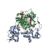
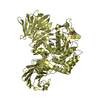
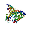
- PDB-1fvh: CRYSTAL STRUCTURE ANALYSIS OF NEURONAL SEC1 FROM THE SQUID L. PEALEI -
+ Open data

- Basic information
| Entry | Database: PDB / ID: 1fvh | ||||||
|---|---|---|---|---|---|---|---|
| Title | CRYSTAL STRUCTURE ANALYSIS OF NEURONAL SEC1 FROM THE SQUID L. PEALEI | ||||||
 Components Components | SEC1 | ||||||
 Keywords Keywords | ENDOCYTOSIS/EXOCYTOSIS / PARALLEL BETA-SHEETS / LEFT-HAND TURN CONNECTION /  HELICAL BUNDLE / ENDOCYTOSIS-EXOCYTOSIS COMPLEX HELICAL BUNDLE / ENDOCYTOSIS-EXOCYTOSIS COMPLEX | ||||||
| Function / homology |  Function and homology information Function and homology information | ||||||
| Biological species |  Loligo pealei (longfin inshore squid) Loligo pealei (longfin inshore squid) | ||||||
| Method | X-RAY DIFFRACTION / SYNCHROTRON / MOLECULAR REPLACEMENT / Resolution: 2.8 Å | ||||||
 Authors Authors | Bracher, A. / Weissenhorn, W. | ||||||
 Citation Citation | Journal: J.Mol.Biol. / Year: 2001 Title: Crystal structures of neuronal squid Sec1 implicate inter-domain hinge movement in the release of t-SNAREs. Authors: Bracher, A. / Weissenhorn, W. #1: Journal: Acta Crystallogr.,Sect.D / Year: 2000Title: Crystallization and preliminary X-ray analysis of squid neuronal Sec1 Authors: Bracher, A. / Dresbach, T. / Betz, H. / Weissenhorn, W. #2: Journal: Structure / Year: 2000Title: The X-ray Crystal Structure of Neuronal Sec1 from Squid Sheds New Light on the Role of this Protein in Exocytosis Authors: Bracher, A. / Perrakis, A. / Dresbach, T. / Betz, H. / Weissenhorn, W. | ||||||
| History |
|
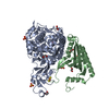
- Structure visualization
| Structure viewer | Molecule: MolmilJmol/JSmol |
|---|
- Downloads & links
-Download
| PDBx/mmCIF format | 1fvh.cif.gz | 121.4 KB | Display | PDBx/mmCIF format |
|---|---|---|---|---|
| PDB format | pdb1fvh.ent.gz | 93.9 KB | Display | PDB format |
| PDBx/mmJSON format | 1fvh.json.gz | Tree view | PDBx/mmJSON format | |
| Others |  Other downloads Other downloads |
-Validation report
| Arichive directory | https://data.pdbj.org/pub/pdb/validation_reports/fv/1fvhftp://data.pdbj.org/pub/pdb/validation_reports/fv/1fvh | HTTPS FTP |
|---|
-Related structure data
| Related structure data | 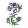 1fvfC 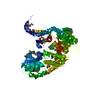 1epuS S: Starting model for refinement C: citing same article ( |
|---|---|
| Similar structure data |


-Links
 PDBj
PDBj
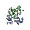
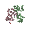
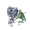
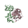
- Assembly
| Deposited unit | 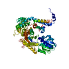
| ||||||||
|---|---|---|---|---|---|---|---|---|---|
| 1 |
| ||||||||
| Unit cell |
|
-Components
| #1: Protein | Mass: 67830.664 Da / Num. of mol.: 1 Source method: isolated from a genetically manipulated source Source: (gene. exp.) Loligo pealei (longfin inshore squid) / Description: LOLIGO, PEALEI, GIANT AXON / Plasmid: PQE30 / Production host:  Escherichia coli (E. coli) / References: UniProt: O62547 Escherichia coli (E. coli) / References: UniProt: O62547 |
|---|---|
| #2: Water | ChemComp-HOH / Water Mass: 18.015 Da / Num. of mol.: 37 / Source method: isolated from a natural source / Formula: H2O Mass: 18.015 Da / Num. of mol.: 37 / Source method: isolated from a natural source / Formula: H2O |
-Experimental details
-Experiment
| Experiment | Method: X-RAY DIFFRACTION / Number of used crystals: 1 |
|---|
- Sample preparation
| Crystal | Density Matthews: 2.94 Å3/Da / Density % sol: 57.8 % | ||||||||||||||||||||||||||||||||||||
|---|---|---|---|---|---|---|---|---|---|---|---|---|---|---|---|---|---|---|---|---|---|---|---|---|---|---|---|---|---|---|---|---|---|---|---|---|---|
| Crystal grow | Temperature: 277 K / Method: batch crystallization / pH: 7.4 Details: Hepes, potassium chloride, dithiothreitol , pH 7.4, BATCH CRYSTALLIZATION, temperature 277K | ||||||||||||||||||||||||||||||||||||
| Crystal grow | *PLUS pH: 5.6 / Method: vapor diffusion, hanging dropDetails: Bracher, A., (2000) Acta Crystallogr., Sect.D, 56, 501. | ||||||||||||||||||||||||||||||||||||
| Components of the solutions | *PLUS
|
-Data collection
| Diffraction | Mean temperature: 100 K |
|---|---|
| Diffraction source | Source: SYNCHROTRON / Site: ESRF  / Beamline: ID14-1 / Wavelength: 0.939 / Beamline: ID14-1 / Wavelength: 0.939 |
| Detector | Type: ADSC QUANTUM 4 / Detector: CCD / Date: Mar 7, 2000 |
| Radiation | Protocol: SINGLE WAVELENGTH / Monochromatic (M) / Laue (L): M / Scattering type: x-ray |
| Radiation wavelength | Wavelength: 0.939 Å / Relative weight: 1 |
| Reflection | Resolution: 2.8→15 Å / Num. all: 17776 / Num. obs: 16849 / % possible obs: 94.8 % / Observed criterion σ(F): 0 / Observed criterion σ(I): 0 / Redundancy: 3.7 % / Biso Wilson estimate: 60.02 Å2 / Rmerge(I) obs: 0.054 / Net I/σ(I): 20.1 |
| Reflection shell | Resolution: 2.8→2.9 Å / Redundancy: 2.9 % / Rmerge(I) obs: 0.287 / Mean I/σ(I) obs: 2.5 / Num. unique all: 1293 / % possible all: 72.6 |
- Processing
| Software |
| ||||||||||||||||||||||||||||||||||||
|---|---|---|---|---|---|---|---|---|---|---|---|---|---|---|---|---|---|---|---|---|---|---|---|---|---|---|---|---|---|---|---|---|---|---|---|---|---|
| Refinement | Method to determine structure: MOLECULAR REPLACEMENT Starting model: PDB ENTRY 1EPU Resolution: 2.8→14.91 Å / Rfactor Rfree error: 0.01 / Isotropic thermal model: restrained / Cross valid method: THROUGHOUT / σ(F): 0 / σ(I): 0 / Stereochemistry target values: Engh & Huber / Details: maximum likelihood function
| ||||||||||||||||||||||||||||||||||||
| Solvent computation | Solvent model: flat model / Bsol: 34.6129 Å2 / ksol: 0.323526 e/Å3 | ||||||||||||||||||||||||||||||||||||
| Displacement parameters | Biso mean: 61.74 Å2
| ||||||||||||||||||||||||||||||||||||
| Refine analyze |
| ||||||||||||||||||||||||||||||||||||
| Refinement step | Cycle: LAST / Resolution: 2.8→14.91 Å
| ||||||||||||||||||||||||||||||||||||
| Refine LS restraints |
| ||||||||||||||||||||||||||||||||||||
| LS refinement shell | Resolution: 2.8→2.9 Å / Rfactor Rfree error: 0.044 / Total num. of bins used: 10
| ||||||||||||||||||||||||||||||||||||
| Xplor file |
| ||||||||||||||||||||||||||||||||||||
| Software | *PLUS Name: CNS / Classification: refinement | ||||||||||||||||||||||||||||||||||||
| Refinement | *PLUS Highest resolution: 2.8 Å / σ(F): 0 / % reflection Rfree: 4.9 % / Rfactor obs: 0.234 | ||||||||||||||||||||||||||||||||||||
| Solvent computation | *PLUS | ||||||||||||||||||||||||||||||||||||
| Displacement parameters | *PLUS | ||||||||||||||||||||||||||||||||||||
| Refine LS restraints | *PLUS
| ||||||||||||||||||||||||||||||||||||
| LS refinement shell | *PLUS Rfactor Rfree: 0.388 / % reflection Rfree: 5.7 % / Rfactor Rwork: 0.41 / Rfactor obs: 0.41 |
