 Movie
Movie Controller

[English] 日本語
 Yorodumi
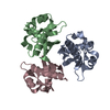
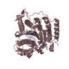
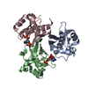
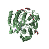
- PDB-1fte: CRYSTAL STRUCTURE OF STREPTOCOCCUS PNEUMONIAE ACYL CARRIER PROTEI... -
+ Open data

- Basic information
| Entry | Database: PDB / ID: 1fte | ||||||
|---|---|---|---|---|---|---|---|
| Title | CRYSTAL STRUCTURE OF STREPTOCOCCUS PNEUMONIAE ACYL CARRIER PROTEIN SYNTHASE (NATIVE 1) | ||||||
 Components Components | ACYL CARRIER PROTEIN SYNTHASE Holo-(acyl-carrier-protein) synthase Holo-(acyl-carrier-protein) synthase | ||||||
 Keywords Keywords | TRANSFERASE / Acyl Carrier Protein Synthase | ||||||
| Function / homology |  Function and homology information Function and homology informationholo-[acyl-carrier-protein] synthase / holo-[acyl-carrier-protein] synthase activity / fatty acid biosynthetic process / magnesium ion binding / cytoplasmSimilarity search - Function | ||||||
| Biological species |   Streptococcus pneumoniae (bacteria) Streptococcus pneumoniae (bacteria) | ||||||
| Method | X-RAY DIFFRACTION / SYNCHROTRON / MAD / Resolution: 2.4 Å | ||||||
 Authors Authors | Chirgadze, N. / Briggs, S. / McAllister, K. / Fischl, A. / Zhao, G. | ||||||
 Citation Citation | Journal: EMBO J. / Year: 2000 Title: Crystal structure of Streptococcus pneumoniae acyl carrier protein synthase: an essential enzyme in bacterial fatty acid biosynthesis. Authors: Chirgadze, N.Y. / Briggs, S.L. / McAllister, K.A. / Fischl, A.S. / Zhao, G. | ||||||
| History |
|
- Structure visualization
| Structure viewer | Molecule: MolmilJmol/JSmol |
|---|
- Downloads & links
-Download
| PDBx/mmCIF format | 1fte.cif.gz | 79.3 KB | Display | PDBx/mmCIF format |
|---|---|---|---|---|
| PDB format | pdb1fte.ent.gz | 60 KB | Display | PDB format |
| PDBx/mmJSON format | 1fte.json.gz | Tree view | PDBx/mmJSON format | |
| Others |  Other downloads Other downloads |
-Validation report
| Arichive directory | https://data.pdbj.org/pub/pdb/validation_reports/ft/1fteftp://data.pdbj.org/pub/pdb/validation_reports/ft/1fte | HTTPS FTP |
|---|
-Related structure data


-Links
 PDBj
PDBj- Assembly
| Deposited unit | 
| ||||||||||
|---|---|---|---|---|---|---|---|---|---|---|---|
| 1 |
| ||||||||||
| Unit cell |
|
-Components
| #1: Protein | Holo-(acyl-carrier-protein) synthase / E.C.2.7.8.7 / ACPS Mass: 13678.506 Da / Num. of mol.: 3 Source method: isolated from a genetically manipulated source Source: (gene. exp.) Streptococcus pneumoniae (bacteria) / Production host: Escherichia coli (E. coli)References: UniProt: P0A2W6, holo-[acyl-carrier-protein] synthase #2: Chemical | Sulfate  Mass: 96.063 Da / Num. of mol.: 2 / Source method: obtained synthetically / Formula: SO4 Mass: 96.063 Da / Num. of mol.: 2 / Source method: obtained synthetically / Formula: SO4#3: Water | ChemComp-HOH / | Water Mass: 18.015 Da / Num. of mol.: 91 / Source method: isolated from a natural source / Formula: H2O Mass: 18.015 Da / Num. of mol.: 91 / Source method: isolated from a natural source / Formula: H2O |
|---|
-Experimental details
-Experiment
| Experiment | Method: X-RAY DIFFRACTION / Number of used crystals: 1 |
|---|
- Sample preparation
| Crystal | Density Matthews: 2.06 Å3/Da / Density % sol: 40.2 % | ||||||||||||||||||||||||||||||||||||||||||||||||
|---|---|---|---|---|---|---|---|---|---|---|---|---|---|---|---|---|---|---|---|---|---|---|---|---|---|---|---|---|---|---|---|---|---|---|---|---|---|---|---|---|---|---|---|---|---|---|---|---|---|
| Crystal grow | *PLUS Temperature: 294 K / pH: 7.1 / Method: vapor diffusion | ||||||||||||||||||||||||||||||||||||||||||||||||
| Components of the solutions | *PLUS
|
-Data collection
| Diffraction | Mean temperature: 100 K |
|---|---|
| Diffraction source | Source: SYNCHROTRON / Site: APS  / Beamline: 17-ID / Wavelength: 1 / Beamline: 17-ID / Wavelength: 1 |
| Detector | Type: MARRESEARCH / Detector: CCD / Date: Apr 29, 1999 |
| Radiation | Protocol: SINGLE WAVELENGTH / Monochromatic (M) / Laue (L): M / Scattering type: x-ray |
| Radiation wavelength | Wavelength: 1 Å / Relative weight: 1 |
| Reflection | Resolution: 2.4→20 Å / Num. all: 14842 / Num. obs: 14842 / % possible obs: 96 % / Observed criterion σ(I): 0 / Redundancy: 4 % / Rmerge(I) obs: 0.049 / Net I/σ(I): 25.6 |
| Reflection shell | Resolution: 2.4→2.49 Å / % possible obs: 84 % / Rmerge(I) obs: 0.23 / Mean I/σ(I) obs: 4.5 / % possible all: 84 |
| Reflection | *PLUS Num. all: 59335 / % possible obs: 96 % / Redundancy: 4 % / Num. measured all: 336325 |
| Reflection shell | *PLUS Rmerge(I) obs: 0.23 |
- Processing
| Software |
| |||||||||||||||||||||||||
|---|---|---|---|---|---|---|---|---|---|---|---|---|---|---|---|---|---|---|---|---|---|---|---|---|---|---|
| Refinement | Method to determine structure: MAD / Resolution: 2.4→20 Å / Cross valid method: THROUGHOUT / σ(F): 0 / σ(I): 0
| |||||||||||||||||||||||||
| Refinement step | Cycle: LAST / Resolution: 2.4→20 Å
| |||||||||||||||||||||||||
| Refine LS restraints |
| |||||||||||||||||||||||||
| LS refinement shell | Resolution: 2.4→2.46 Å / Total num. of bins used: 13
| |||||||||||||||||||||||||
| Software | *PLUS Name: CNX / Classification: refinement | |||||||||||||||||||||||||
| Refinement | *PLUS Lowest resolution: 20 Å / σ(F): 0 / % reflection Rfree: 4.9 % / Rfactor obs: 0.229 | |||||||||||||||||||||||||
| Solvent computation | *PLUS | |||||||||||||||||||||||||
| Displacement parameters | *PLUS | |||||||||||||||||||||||||
| Refine LS restraints | *PLUS
| |||||||||||||||||||||||||
| LS refinement shell | *PLUS % reflection Rfree: 4.9 % / Rfactor Rwork: 0.309 |
