 Movie
Movie Controller

[English] 日本語
 Yorodumi
- PDB-1fpz: CRYSTAL STRUCTURE ANALYSIS OF KINASE ASSOCIATED PHOSPHATASE (KAP)... -
+ Open data

- Basic information
| Entry | Database: PDB / ID: 1fpz | ||||||
|---|---|---|---|---|---|---|---|
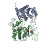
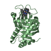
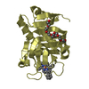
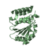
| Title | CRYSTAL STRUCTURE ANALYSIS OF KINASE ASSOCIATED PHOSPHATASE (KAP) WITH A SUBSTITUTION OF THE CATALYTIC SITE CYSTEINE (CYS140) TO A SERINE | ||||||
 Components Components | CYCLIN-DEPENDENT KINASE INHIBITOR 3 Cyclin-dependent kinase inhibitor protein Cyclin-dependent kinase inhibitor protein | ||||||
 Keywords Keywords | HYDROLASE / alpha-beta sandwich | ||||||
| Function / homology |  Function and homology information Function and homology informationprotein tyrosine/serine/threonine phosphatase activity / myosin phosphatase activity / protein serine/threonine phosphatase activity / protein-serine/threonine phosphatase / regulation of cyclin-dependent protein serine/threonine kinase activity / dephosphorylation / protein-tyrosine-phosphatase / protein tyrosine phosphatase activity / G1/S transition of mitotic cell cycle / regulation of cell cycle ...protein tyrosine/serine/threonine phosphatase activity / myosin phosphatase activity / protein serine/threonine phosphatase activity / protein-serine/threonine phosphatase / regulation of cyclin-dependent protein serine/threonine kinase activity / dephosphorylation / protein-tyrosine-phosphatase / protein tyrosine phosphatase activity / G1/S transition of mitotic cell cycle / regulation of cell cycle / negative regulation of cell population proliferation / perinuclear region of cytoplasm / nucleus / cytosol / cytoplasmSimilarity search - Function | ||||||
| Biological species |  Homo sapiens (human) Homo sapiens (human) | ||||||
| Method | X-RAY DIFFRACTION / SYNCHROTRON / Resolution: 2 Å | ||||||
 Authors Authors | Song, H. / Hanlon, N. / Brown, N.R. / Noble, M.E.M. / Johnson, L.N. / Barford, D. | ||||||
 Citation Citation | Journal: Mol.Cell / Year: 2001 Title: Phosphoprotein-protein interactions revealed by the crystal structure of kinase-associated phosphatase in complex with phosphoCDK2. Authors: Song, H. / Hanlon, N. / Brown, N.R. / Noble, M.E. / Johnson, L.N. / Barford, D. #1: Journal: Cell(Cambridge,Mass.) / Year: 1993Title: Cdi1, a human G1 and S phase protein phosphatase that associates with Cdk2 Authors: Gyruris, J. / Golemis, E. / Chertkov, H. / Brent, R. #2: Journal: Cell(Cambridge,Mass.) / Year: 1993Title: The p21Cdk-interacting protein Cip1 is a potent inhibitor of G1 cyclin-dependent kinases Authors: Harper, J.W. / Adami, G.R. / Wei, N. / Keyomarsi, K. / Elledge, S.J. | ||||||
| History |
|
- Structure visualization
| Structure viewer | Molecule: MolmilJmol/JSmol |
|---|
- Downloads & links
-Download
| PDBx/mmCIF format | 1fpz.cif.gz | 230.6 KB | Display | PDBx/mmCIF format |
|---|---|---|---|---|
| PDB format | pdb1fpz.ent.gz | 186.2 KB | Display | PDB format |
| PDBx/mmJSON format | 1fpz.json.gz | Tree view | PDBx/mmJSON format | |
| Others |  Other downloads Other downloads |
-Validation report
| Arichive directory | https://data.pdbj.org/pub/pdb/validation_reports/fp/1fpzftp://data.pdbj.org/pub/pdb/validation_reports/fp/1fpz | HTTPS FTP |
|---|
-Related structure data


-Links
 PDBj
PDBj


- Assembly
| Deposited unit | 
| ||||||||
|---|---|---|---|---|---|---|---|---|---|
| 1 | 
| ||||||||
| 2 | 
| ||||||||
| 3 | 
| ||||||||
| 4 | 
| ||||||||
| 5 | 
| ||||||||
| 6 | 
| ||||||||
| Unit cell |
|
-Components
| #1: Protein | Cyclin-dependent kinase inhibitor protein Mass: 23819.057 Da / Num. of mol.: 6 / Mutation: C140S Source method: isolated from a genetically manipulated source Source: (gene. exp.) Homo sapiens (human) / Description: HOMO SAPIENS / Production host:  Escherichia coli (E. coli) / References: UniProt: Q16667, protein-tyrosine-phosphatase Escherichia coli (E. coli) / References: UniProt: Q16667, protein-tyrosine-phosphatase#2: Chemical | ChemComp-SO4 / Sulfate  Mass: 96.063 Da / Num. of mol.: 6 / Source method: obtained synthetically / Formula: SO4 Mass: 96.063 Da / Num. of mol.: 6 / Source method: obtained synthetically / Formula: SO4#3: Water | ChemComp-HOH / | Water Mass: 18.015 Da / Num. of mol.: 690 / Source method: isolated from a natural source / Formula: H2O Mass: 18.015 Da / Num. of mol.: 690 / Source method: isolated from a natural source / Formula: H2O |
|---|
-Experimental details
-Experiment
| Experiment | Method: X-RAY DIFFRACTION / Number of used crystals: 1 |
|---|
- Sample preparation
| Crystal | Density Matthews: 2.46 Å3/Da / Density % sol: 50.09 % | ||||||||||||||||||||||||||||||
|---|---|---|---|---|---|---|---|---|---|---|---|---|---|---|---|---|---|---|---|---|---|---|---|---|---|---|---|---|---|---|---|
| Crystal grow | Temperature: 277 K / Method: vapor diffusion, hanging drop / pH: 5 Details: PEG 8000, ammonium sulphate, sodium acetate, pH 5.0, VAPOR DIFFUSION, HANGING DROP, temperature 277K | ||||||||||||||||||||||||||||||
| Crystal grow | *PLUS Temperature: 4 ℃ / Details: Hanlon, N., (1998) Protein Sci., 7, 508. | ||||||||||||||||||||||||||||||
| Components of the solutions | *PLUS
|
-Data collection
| Diffraction | Mean temperature: 100 K |
|---|---|
| Diffraction source | Source: SYNCHROTRON / Site: SRS  / Beamline: PX9.6 / Wavelength: 0.87 / Beamline: PX9.6 / Wavelength: 0.87 |
| Detector | Type: ADSC / Detector: CCD / Date: Jan 1, 2000 |
| Radiation | Protocol: SINGLE WAVELENGTH / Monochromatic (M) / Laue (L): M / Scattering type: x-ray |
| Radiation wavelength | Wavelength: 0.87 Å / Relative weight: 1 |
| Reflection | Resolution: 2→50 Å / Num. obs: 90055 / % possible obs: 96.7 % / Observed criterion σ(F): 2 / Observed criterion σ(I): 2 / Redundancy: 3.6 % / Biso Wilson estimate: 23.6 Å2 / Rmerge(I) obs: 0.06 / Net I/σ(I): 9.3 |
| Reflection shell | Resolution: 2→2.12 Å / Rmerge(I) obs: 0.38 / % possible all: 91.6 |
| Reflection | *PLUS Num. measured all: 319770 |
| Reflection shell | *PLUS % possible obs: 91.6 % / Mean I/σ(I) obs: 1.9 |
- Processing
| Software |
| ||||||||||||||||||||
|---|---|---|---|---|---|---|---|---|---|---|---|---|---|---|---|---|---|---|---|---|---|
| Refinement | Resolution: 2→20 Å / Cross valid method: THROUGHOUT / σ(F): 0 / σ(I): 0 / Stereochemistry target values: Engh & Huber
| ||||||||||||||||||||
| Refinement step | Cycle: LAST / Resolution: 2→20 Å
| ||||||||||||||||||||
| Refine LS restraints |
| ||||||||||||||||||||
| Software | *PLUS Name: CNS / Classification: refinement | ||||||||||||||||||||
| Refinement | *PLUS Highest resolution: 2 Å / Lowest resolution: 20 Å / σ(F): 0 / % reflection Rfree: 10 % / Rfactor obs: 0.202 | ||||||||||||||||||||
| Solvent computation | *PLUS | ||||||||||||||||||||
| Displacement parameters | *PLUS |
