 Movie
Movie Controller

[English] 日本語
 Yorodumi
- PDB-1fo0: MURINE ALLOREACTIVE SCFV TCR-PEPTIDE-MHC CLASS I MOLECULE COMPLEX -
+ Open data

- Basic information
| Entry | Database: PDB / ID: 1fo0 | ||||||
|---|---|---|---|---|---|---|---|
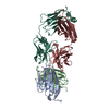
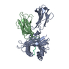
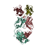
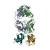
| Title | MURINE ALLOREACTIVE SCFV TCR-PEPTIDE-MHC CLASS I MOLECULE COMPLEX | ||||||
 Components Components |
| ||||||
 Keywords Keywords |  IMMUNE SYSTEM / T CELL RECEPTOR / CLASS I MHC / H-2KB / TCR-PMHC COMPLEX IMMUNE SYSTEM / T CELL RECEPTOR / CLASS I MHC / H-2KB / TCR-PMHC COMPLEX | ||||||
| Function / homology |  Function and homology information Function and homology informationEndosomal/Vacuolar pathway / DAP12 interactions / Antigen Presentation: Folding, assembly and peptide loading of class I MHC / ER-Phagosome pathway / DAP12 signaling / Immunoregulatory interactions between a Lymphoid and a non-Lymphoid cell / T cell receptor complex / regulation of membrane depolarization / antigen processing and presentation of exogenous peptide antigen via MHC class I / inner ear development ...Endosomal/Vacuolar pathway / DAP12 interactions / Antigen Presentation: Folding, assembly and peptide loading of class I MHC / ER-Phagosome pathway / DAP12 signaling / Immunoregulatory interactions between a Lymphoid and a non-Lymphoid cell / T cell receptor complex / regulation of membrane depolarization / antigen processing and presentation of exogenous peptide antigen via MHC class I / inner ear development / antigen processing and presentation of endogenous peptide antigen via MHC class I via ER pathway, TAP-dependent / cellular defense response / beta-2-microglobulin binding / positive regulation of phosphorylation / Neutrophil degranulation / antigen processing and presentation of endogenous peptide antigen via MHC class I via ER pathway, TAP-independent / antigen processing and presentation of endogenous peptide antigen via MHC class Ib / lumenal side of endoplasmic reticulum membrane / peptide binding / cellular response to iron(III) ion / antigen processing and presentation of exogenous protein antigen via MHC class Ib, TAP-dependent / negative regulation of forebrain neuron differentiation / regulation of erythrocyte differentiation / peptide antigen assembly with MHC class I protein complex / response to molecule of bacterial origin / regulation of iron ion transport / MHC class I peptide loading complex / HFE-transferrin receptor complex / T cell mediated cytotoxicity / cellular response to iron ion / antigen processing and presentation of endogenous peptide antigen via MHC class I / positive regulation of T cell cytokine production / MHC class I protein complex / multicellular organismal-level iron ion homeostasis / negative regulation of neurogenesis / peptide antigen assembly with MHC class II protein complex / positive regulation of receptor-mediated endocytosis / positive regulation of T cell mediated cytotoxicity / MHC class II protein complex / cellular response to nicotine / phagocytic vesicle membrane / peptide antigen binding / positive regulation of cellular senescence / antigen processing and presentation of exogenous peptide antigen via MHC class II / negative regulation of epithelial cell proliferation / positive regulation of immune response / antimicrobial humoral immune response mediated by antimicrobial peptide / sensory perception of smell / positive regulation of T cell activation / negative regulation of neuron projection development / MHC class II protein complex binding / late endosome membrane / T cell differentiation in thymus / iron ion transport / protein refolding / antibacterial humoral response / protein homotetramerization / intracellular iron ion homeostasis / adaptive immune response / cellular response to lipopolysaccharide / defense response to Gram-negative bacterium / amyloid fibril formation / learning or memory / defense response to Gram-positive bacterium / defense response to bacterium / immune response / lysosomal membrane / external side of plasma membrane / signaling receptor binding / innate immune response / protein-containing complex binding / structural molecule activity / Golgi apparatus / protein homodimerization activity / extracellular space / identical protein binding / plasma membrane / cytosol / cytoplasmSimilarity search - Function | ||||||
| Biological species |  Mus musculus (house mouse) Mus musculus (house mouse) | ||||||
| Method | X-RAY DIFFRACTION / SYNCHROTRON / MOLECULAR REPLACEMENT / Resolution: 2.5 Å | ||||||
 Authors Authors | Reiser, J.B. / Darnault, C. / Guimezanes, A. / Gregoire, C. / Mosser, T. / Schmitt-Verhulst, A.-M. / Fontecilla-Camps, J.C. / Malissen, B. / Housset, D. / Mazza, G. | ||||||
 Citation Citation | Journal: Nat.Immunol. / Year: 2000 Title: Crystal structure of a T cell receptor bound to an allogeneic MHC molecule. Authors: Reiser, J.B. / Darnault, C. / Guimezanes, A. / Gregoire, C. / Mosser, T. / Schmitt-Verhulst, A.-M. / Fontecilla-Camps, J.C. / Malissen, B. / Housset, D. / Mazza, G. #1: Journal: Embo J. / Year: 1997Title: The Three-Dimensional Structure of a T Cell Receptor Valpha-Vbeta Heterodimer Reveals a Novel Arrangement of the Vbeta Domain Authors: Housset, D. / Mazza, G. / Gregoire, C. / Piras, C. / Malissen, B. / Fontecilla-Camps, J.C. #2: Journal: Proc.Natl.Acad.Sci.USA / Year: 1995Title: Crystal Structure of an H-2Kb-Ovalbumin Peptide Com Reveals the Interplay of Primary and Secondary Anchor Positions in the Major Histocompatibility Complex Binding Groove Authors: Fremont, D.H. / Stura, E.A. / Matsumura, M. / Peterson, P.A. / Wilson, I.A. #3: Journal: Science / Year: 1998Title: Structural Basis of Plasticity in T Cell Receptor Recognition of a Self Peptide-Mhc Antigen Authors: Garcia, K.C. / Degano, M. / Pease, L.R. / Huang, M. / Peterson, P.A. / Teyton, L. / Wilson, I.A. #4: Journal: Nature / Year: 1996Title: Structure of the Complex between Human T Cell Receptor, Viral Peptide and Hla-A2 Authors: Garboczi, D.N. / Ghosh, P. / Utz, U. / Fan, Q.R. / Bidisson, W.E. / Wiley, D. #5: Journal: Immunity / Year: 1998Title: Two Human T Cell Receptors Bind in a Similar Mode To the Hla-A2/Tax Peptide Complex Using Different Tcr Amino-acids Authors: Ding, Y.H. / Smith, K.J. / Garboczi, D.N. / Utz, U. / Bidisson, W.E. / Wiley, D.C. #6: Journal: Int.Immunol. / Year: 1991Title: Each of Two Productive T Cell Receptor Alpha-Gene Rearrangements Found in Both the A10 and Bm3.3 Cell Clones Give Rise to an Alpha Chain which Can Contribute to the Constitution of a Surface- ...Title: Each of Two Productive T Cell Receptor Alpha-Gene Rearrangements Found in Both the A10 and Bm3.3 Cell Clones Give Rise to an Alpha Chain which Can Contribute to the Constitution of a Surface-Expressed Alpha-Beta Dimer Authors: Couez, D. / Malissen, M. / Buferne, M. / Schmitt-Verhulst, A.-M. / Malissen, B. | ||||||
| History |
|
- Structure visualization
| Structure viewer | Molecule: MolmilJmol/JSmol |
|---|
- Downloads & links
-Download
| PDBx/mmCIF format | 1fo0.cif.gz | 141.5 KB | Display | PDBx/mmCIF format |
|---|---|---|---|---|
| PDB format | pdb1fo0.ent.gz | 109.9 KB | Display | PDB format |
| PDBx/mmJSON format | 1fo0.json.gz | Tree view | PDBx/mmJSON format | |
| Others |  Other downloads Other downloads |
-Validation report
| Arichive directory | https://data.pdbj.org/pub/pdb/validation_reports/fo/1fo0ftp://data.pdbj.org/pub/pdb/validation_reports/fo/1fo0 | HTTPS FTP |
|---|
-Related structure data


-Links
 PDBj
PDBj


- Assembly
| Deposited unit | 
| ||||||||
|---|---|---|---|---|---|---|---|---|---|
| 1 |
| ||||||||
| Unit cell |
|
-Components
-Protein , 2 types, 2 molecules HL
| #1: Protein | Mass: 31908.629 Da / Num. of mol.: 1 / Fragment: EXTRACELLULAR DOMAINS (ALPHA1, ALPHA2, ALPHA3) Source method: isolated from a genetically manipulated source Source: (gene. exp.) Mus musculus (house mouse) / Production host:  Escherichia coli (E. coli) / References: UniProt: P01901 Escherichia coli (E. coli) / References: UniProt: P01901 |
|---|---|
| #2: Protein | Mass: 11704.359 Da / Num. of mol.: 1 Source method: isolated from a genetically manipulated source Source: (gene. exp.) Mus musculus (house mouse) / Production host: Escherichia coli (E. coli) / References: UniProt: P01887 |
-PROTEIN (BM3.3 T CELL RECEPTOR ... , 2 types, 2 molecules AB
| #4: Protein | Mass: 12945.639 Da / Num. of mol.: 1 / Fragment: FV FRAGMENT, VARIABLE DOMAIN Source method: isolated from a genetically manipulated source Source: (gene. exp.) Mus musculus (house mouse) / Cell (production host): MYELOMA CELLS / Production host: Mus musculus (house mouse) / References: GenBank: 201157, UniProt: Q5R1F1*PLUS |
|---|---|
| #5: Protein | Mass: 12965.832 Da / Num. of mol.: 1 / Fragment: FV FRAGMENT, VARIABLE DOMAIN Source method: isolated from a genetically manipulated source Source: (gene. exp.) Mus musculus (house mouse) / Cell (production host): MYELOMA CELLS / Production host: Mus musculus (house mouse) / References: GenBank: 554307, UniProt: P04214*PLUS |
-Protein/peptide / Non-polymers , 2 types, 192 molecules P
| #3: Protein/peptide | Mass: 983.076 Da / Num. of mol.: 1 / Source method: obtained synthetically / Details: SEQUENCE NATURALLY OCCURS IN MUS MUCULUS / References: UniProt: Q8CDD8*PLUS |
|---|---|
| #6: Water | ChemComp-HOH / WaterMass: 18.015 Da / Num. of mol.: 191 / Source method: isolated from a natural source / Formula: H2O |
-Experimental details
-Experiment
| Experiment | Method: X-RAY DIFFRACTION / Number of used crystals: 1 |
|---|
- Sample preparation
| Crystal | Density Matthews: 3.2 Å3/Da / Density % sol: 62 % / Description: RESOLUTION RANGE USED FOR MR 15.0-3.5 A | |||||||||||||||||||||||||
|---|---|---|---|---|---|---|---|---|---|---|---|---|---|---|---|---|---|---|---|---|---|---|---|---|---|---|
| Crystal grow | Method: vapor diffusion / pH: 7 Details: PEG 6000 10% HEPES 0.1M PH 7.0 MGAC 0.25 M NACL 0.25M, pH 7.00, VAPOR DIFFUSION | |||||||||||||||||||||||||
| Crystal grow | *PLUS Temperature: 4 ℃ / Method: vapor diffusion, hanging drop | |||||||||||||||||||||||||
| Components of the solutions | *PLUS
|
-Data collection
| Diffraction | Mean temperature: 110 K |
|---|---|
| Diffraction source | Source: SYNCHROTRON / Site: ESRF  / Beamline: ID14-3 / Wavelength: 0.931 / Beamline: ID14-3 / Wavelength: 0.931 |
| Detector | Type: MARRESEARCH / Detector: CCD / Date: Dec 8, 1999 |
| Radiation | Protocol: SINGLE WAVELENGTH / Monochromatic (M) / Laue (L): M / Scattering type: x-ray |
| Radiation wavelength | Wavelength: 0.931 Å / Relative weight: 1 |
| Reflection | Resolution: 2.5→23.1 Å / Num. obs: 33614 / % possible obs: 99.9 % / Observed criterion σ(I): 0 / Redundancy: 7.2 % / Biso Wilson estimate: 49.87 Å2 / Rsym value: 0.081 / Net I/σ(I): 8.3 |
| Reflection shell | Resolution: 2.5→2.64 Å / Redundancy: 7.1 % / Mean I/σ(I) obs: 1.9 / Rsym value: 0.375 / % possible all: 100 |
| Reflection | *PLUS Rmerge(I) obs: 0.081 |
| Reflection shell | *PLUS % possible obs: 100 % / Rmerge(I) obs: 0.375 |
- Processing
| Software |
| ||||||||||||||||||||||||||||||||||||||||||||||||||||||||||||||||||||||||||||||||||||
|---|---|---|---|---|---|---|---|---|---|---|---|---|---|---|---|---|---|---|---|---|---|---|---|---|---|---|---|---|---|---|---|---|---|---|---|---|---|---|---|---|---|---|---|---|---|---|---|---|---|---|---|---|---|---|---|---|---|---|---|---|---|---|---|---|---|---|---|---|---|---|---|---|---|---|---|---|---|---|---|---|---|---|---|---|---|
| Refinement | Method to determine structure: MOLECULAR REPLACEMENT Starting model: PDB ENTRIES 1KB5, 1VAC Resolution: 2.5→12 Å / Cross valid method: THROUGHOUT / σ(F): 0 / Details: BULK SOLVENT CORRECTION USED R V
| ||||||||||||||||||||||||||||||||||||||||||||||||||||||||||||||||||||||||||||||||||||
| Displacement parameters | Biso mean: 57.93 Å2 | ||||||||||||||||||||||||||||||||||||||||||||||||||||||||||||||||||||||||||||||||||||
| Refinement step | Cycle: LAST / Resolution: 2.5→12 Å
| ||||||||||||||||||||||||||||||||||||||||||||||||||||||||||||||||||||||||||||||||||||
| Refine LS restraints |
|
