 Movie
Movie Controller


+ Open data

- Basic information
| Entry | Database: PDB / ID: 1fkk | ||||||
|---|---|---|---|---|---|---|---|
| Title | ATOMIC STRUCTURE OF FKBP12, AN IMMUNOPHILIN BINDING PROTEIN | ||||||
 Components Components | FK506 BINDING PROTEIN FKBP FKBP | ||||||
 Keywords Keywords | ROTAMASE / FK506 BINDING PROTEIN / FKBP12 / CIS-TRANS PROLYL-ISOMERASE | ||||||
| Function / homology |  Function and homology information Function and homology informationmTORC1-mediated signalling / TGF-beta receptor signaling activates SMADs / regulation of activin receptor signaling pathway / Calcineurin activates NFAT / cytoplasmic side of membrane / activin binding / heart trabecula formation / ryanodine receptor complex / negative regulation of release of sequestered calcium ion into cytosol / regulation of amyloid precursor protein catabolic process ...mTORC1-mediated signalling / TGF-beta receptor signaling activates SMADs / regulation of activin receptor signaling pathway / Calcineurin activates NFAT / cytoplasmic side of membrane / activin binding / heart trabecula formation / ryanodine receptor complex / negative regulation of release of sequestered calcium ion into cytosol / regulation of amyloid precursor protein catabolic process / response to caffeine / ventricular cardiac muscle tissue morphogenesis / negative regulation of phosphoprotein phosphatase activity / FK506 binding / SMAD binding / negative regulation of ryanodine-sensitive calcium-release channel activity / regulation of immune response / calcium channel inhibitor activity / protein peptidyl-prolyl isomerization / supramolecular fiber organization / T cell proliferation / heart morphogenesis / release of sequestered calcium ion into cytosol / regulation of ryanodine-sensitive calcium-release channel activity / sarcoplasmic reticulum membrane / T cell activation / negative regulation of protein phosphorylation / sarcoplasmic reticulum / peptidylprolyl isomerase / positive regulation of protein ubiquitination / peptidyl-prolyl cis-trans isomerase activity / muscle contraction / cytokine-mediated signaling pathway / Z disc / regulation of protein localization / positive regulation of protein binding / positive regulation of canonical NF-kappaB signal transduction / transmembrane transporter binding / amyloid fibril formation / protein homodimerization activity / extracellular exosome / membrane / cytosol / cytoplasmSimilarity search - Function | ||||||
| Biological species |  Bos taurus (cattle) Bos taurus (cattle) | ||||||
| Method | X-RAY DIFFRACTION / Resolution: 2.2 Å | ||||||
 Authors Authors | Wilson, K.P. / Sintchak, M.D. / Thomson, J.A. / Navia, M.A. | ||||||
 Citation Citation | Journal: Acta Crystallogr.,Sect.D / Year: 1995 Title: Comparative X-ray structures of the major binding protein for the immunosuppressant FK506 (tacrolimus) in unliganded form and in complex with FK506 and rapamycin. Authors: Wilson, K.P. / Yamashita, M.M. / Sintchak, M.D. / Rotstein, S.H. / Murcko, M.A. / Boger, J. / Thomson, J.A. / Fitzgibbon, M.J. / Black, J.R. / Navia, M.A. #1: Journal: Cell(Cambridge,Mass.) / Year: 1995Title: X-Ray Structure of Calcineurin Inhibited by the Immunophilin-Immunosuppressant Fkbp12-Fk506 Complex Authors: Griffith, J.P. / Kim, J.L. / Kim, E.E. / Sintchak, M.D. / Thomson, J.A. / Fitzgibbon, M.J. / Fleming, M.A. / Caron, P.R. / Hsiao, K. / Navia, M.A. #2: Journal: J.Biol.Chem. / Year: 1993Title: Improved Calcineurin Inhibition by Yeast Fkbp12-Drug Complexes Authors: Rotonda, J. / Burbaum, J.J. / Chan, H.K. / Marcy, A.I. / Becker, J.W. #3: Journal: J.Mol.Biol. / Year: 1993Title: Fk-506-Binding Protein: Three-Dimensional Structure of the Complex with the Antagonist L-685,818 Authors: Becker, J.W. / Rotonda, J. / Mckeever, B.M. / Chan, H.K. / Marcy, A.I. / Wiederrecht, G. / Hermes, J.D. / Springer, J.P. #4: Journal: J.Mol.Biol. / Year: 1993Title: Atomic Structures of Human Immunophilin Fkbp12 Complexes with Fk506 and Rapamycin Authors: Van Duyne, G.D. / Standaert, R.F. / Karplus, P.A. / Schreiber, S.L. / Clardy, J. #5: Journal: J.Am.Chem.Soc. / Year: 1991Title: Atomic Structure of the Rapamycin Human Immunophilin Fkbp-12 Complex Authors: Van Duyne, G.D. / Standaert, R.F. / Schreiber, S.L. / Clardy, J. #6: Journal: Science / Year: 1991Title: Atomic Structure of Fkbp-Fk506, an Immunophilin-Immunosuppressant Complex Authors: Van Duyne, G.D. / Standaert, R.F. / Karplus, P.A. / Schreiber, S.L. / Clardy, J. | ||||||
| History |
|
- Structure visualization
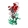
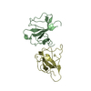
| Structure viewer | Molecule: MolmilJmol/JSmol |
|---|
- Downloads & links
-Download
| PDBx/mmCIF format | 1fkk.cif.gz | 34.6 KB | Display | PDBx/mmCIF format |
|---|---|---|---|---|
| PDB format | pdb1fkk.ent.gz | 23.8 KB | Display | PDB format |
| PDBx/mmJSON format | 1fkk.json.gz | Tree view | PDBx/mmJSON format | |
| Others |  Other downloads Other downloads |
-Validation report
| Arichive directory | https://data.pdbj.org/pub/pdb/validation_reports/fk/1fkkftp://data.pdbj.org/pub/pdb/validation_reports/fk/1fkk | HTTPS FTP |
|---|
-Related structure data


-Links
 PDBj
PDBj

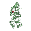
- Assembly
| Deposited unit | 
| ||||||||
|---|---|---|---|---|---|---|---|---|---|
| 1 | 
| ||||||||
| Unit cell |
|
-Components
| #1: Protein | FKBP / FKBP12 Mass: 11794.425 Da / Num. of mol.: 1 / Source method: isolated from a natural source / Details: MR 12,000 DALTONS / Source: (natural) Bos taurus (cattle) / Organ: THYMUS / References: UniProt: P18203, peptidylprolyl isomerase |
|---|---|
| #2: Chemical | ChemComp-SO4 / Sulfate  Mass: 96.063 Da / Num. of mol.: 1 / Source method: obtained synthetically / Formula: SO4 Mass: 96.063 Da / Num. of mol.: 1 / Source method: obtained synthetically / Formula: SO4 |
| #3: Water | ChemComp-HOH / Water Mass: 18.015 Da / Num. of mol.: 67 / Source method: isolated from a natural source / Formula: H2O Mass: 18.015 Da / Num. of mol.: 67 / Source method: isolated from a natural source / Formula: H2O |
-Experimental details
-Experiment
| Experiment | Method: X-RAY DIFFRACTION |
|---|
- Sample preparation
| Crystal | Density Matthews: 2.49 Å3/Da / Density % sol: 50.61 % | ||||||||||||||||||||
|---|---|---|---|---|---|---|---|---|---|---|---|---|---|---|---|---|---|---|---|---|---|
| Crystal grow | *PLUS pH: 7.5 / Method: vapor diffusion | ||||||||||||||||||||
| Components of the solutions | *PLUS
|
-Data collection
| Diffraction source | Wavelength: 1.54 |
|---|---|
| Detector | Detector: IMAGE PLATE |
| Radiation | Monochromatic (M) / Laue (L): M / Scattering type: x-ray |
| Radiation wavelength | Wavelength: 1.54 Å / Relative weight: 1 |
| Reflection | *PLUS Highest resolution: 2.3 Å / Num. obs: 3977 / % possible obs: 68 % / Num. measured all: 10574 / Rmerge(I) obs: 0.049 |
- Processing
| Software |
| ||||||||||||||||||||||||||||||||||||||||||||||||||||||||||||
|---|---|---|---|---|---|---|---|---|---|---|---|---|---|---|---|---|---|---|---|---|---|---|---|---|---|---|---|---|---|---|---|---|---|---|---|---|---|---|---|---|---|---|---|---|---|---|---|---|---|---|---|---|---|---|---|---|---|---|---|---|---|
| Refinement | Resolution: 2.2→7 Å / σ(F): 1 /
| ||||||||||||||||||||||||||||||||||||||||||||||||||||||||||||
| Refinement step | Cycle: LAST / Resolution: 2.2→7 Å
| ||||||||||||||||||||||||||||||||||||||||||||||||||||||||||||
| Refine LS restraints |
| ||||||||||||||||||||||||||||||||||||||||||||||||||||||||||||
| Software | *PLUS Name: X-PLOR / Classification: refinement | ||||||||||||||||||||||||||||||||||||||||||||||||||||||||||||
| Refinement | *PLUS Highest resolution: 2.3 Å | ||||||||||||||||||||||||||||||||||||||||||||||||||||||||||||
| Solvent computation | *PLUS | ||||||||||||||||||||||||||||||||||||||||||||||||||||||||||||
| Displacement parameters | *PLUS | ||||||||||||||||||||||||||||||||||||||||||||||||||||||||||||
| Refine LS restraints | *PLUS Type: x_improper_angle_deg / Dev ideal: 1.9 |
