 Movie
Movie Controller

[English] 日本語
 Yorodumi
- PDB-1fj8: THE STRUCTURE OF BETA-KETOACYL-[ACYL CARRIER PROTEIN] SYNTHASE I ... -
+ Open data

- Basic information
| Entry | Database: PDB / ID: 1fj8 | ||||||
|---|---|---|---|---|---|---|---|
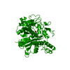
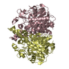
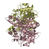
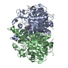
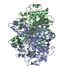
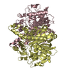
| Title | THE STRUCTURE OF BETA-KETOACYL-[ACYL CARRIER PROTEIN] SYNTHASE I IN COMPLEX WITH CERULENIN, IMPLICATIONS FOR DRUG DESIGN | ||||||
 Components Components | BETA-KETOACYL-[ACYL CARRIER PROTEIN] SYNTHASE I | ||||||
 Keywords Keywords |  TRANSFERASE / CONDENSING ENZYMES / FATTY ACID ELONGATION / CERULENIN / DRUG DESIGN TRANSFERASE / CONDENSING ENZYMES / FATTY ACID ELONGATION / CERULENIN / DRUG DESIGN | ||||||
| Function / homology |  Function and homology information Function and homology informationmonounsaturated fatty acid biosynthetic process / beta-ketoacyl-[acyl-carrier-protein] synthase I / 3-oxoacyl-[acyl-carrier-protein] synthase activity / fatty acid biosynthetic process / cytosolSimilarity search - Function | ||||||
| Biological species |  Escherichia coli (E. coli) Escherichia coli (E. coli) | ||||||
| Method | X-RAY DIFFRACTION / Resolution: 2.27 Å | ||||||
 Authors Authors | Price, A.C. / Choi, K. / Heath, R.J. / Li, Z. / White, S.W. / Rock, C.O. | ||||||
 Citation Citation | Journal: J.Biol.Chem. / Year: 2001 Title: Inhibition of beta-ketoacyl-acyl carrier protein synthases by thiolactomycin and cerulenin. Structure and mechanism. Authors: Price, A.C. / Choi, K.H. / Heath, R.J. / Li, Z. / White, S.W. / Rock, C.O. | ||||||
| History |
|
- Structure visualization
| Structure viewer | Molecule: MolmilJmol/JSmol |
|---|
- Downloads & links
-Download
| PDBx/mmCIF format | 1fj8.cif.gz | 291.1 KB | Display | PDBx/mmCIF format |
|---|---|---|---|---|
| PDB format | pdb1fj8.ent.gz | 246.3 KB | Display | PDB format |
| PDBx/mmJSON format | 1fj8.json.gz | Tree view | PDBx/mmJSON format | |
| Others |  Other downloads Other downloads |
-Validation report
| Arichive directory | https://data.pdbj.org/pub/pdb/validation_reports/fj/1fj8ftp://data.pdbj.org/pub/pdb/validation_reports/fj/1fj8 | HTTPS FTP |
|---|
-Related structure data


-Links
 PDBj
PDBj

- Assembly
| Deposited unit | 
| ||||||||
|---|---|---|---|---|---|---|---|---|---|
| 1 | 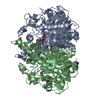
| ||||||||
| 2 | 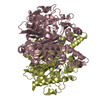
| ||||||||
| Unit cell |
| ||||||||
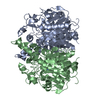
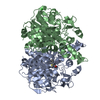
| Details | The biological assembly is a dimer constructed from chain A and chain B. / The biological assembly is a dimer constructed from chain C and chain D. |
-Components
| #1: Protein | Mass: 42656.203 Da / Num. of mol.: 4 Source method: isolated from a genetically manipulated source Source: (gene. exp.) Escherichia coli (E. coli) / Plasmid: PET15B / Production host: Escherichia coli (E. coli)References: UniProt: P0A953, beta-ketoacyl-[acyl-carrier-protein] synthase I #2: Chemical | ChemComp-CER / ( Cerulenin  Mass: 225.284 Da / Num. of mol.: 4 / Source method: obtained synthetically / Formula: C12H19NO3 / Comment: antifungal, antibiotic*YM Mass: 225.284 Da / Num. of mol.: 4 / Source method: obtained synthetically / Formula: C12H19NO3 / Comment: antifungal, antibiotic*YM#3: Water | ChemComp-HOH / | Water Mass: 18.015 Da / Num. of mol.: 239 / Source method: isolated from a natural source / Formula: H2O Mass: 18.015 Da / Num. of mol.: 239 / Source method: isolated from a natural source / Formula: H2O |
|---|
-Experimental details
-Experiment
| Experiment | Method: X-RAY DIFFRACTION / Number of used crystals: 1 |
|---|
- Sample preparation
| Crystal | Density Matthews: 2.57 Å3/Da / Density % sol: 52.14 % | |||||||||||||||||||||||||
|---|---|---|---|---|---|---|---|---|---|---|---|---|---|---|---|---|---|---|---|---|---|---|---|---|---|---|
| Crystal grow | Temperature: 291 K / Method: vapor diffusion, hanging drop / pH: 6.5 Details: ammonium sulfate, PEG 400, Tris, pH 6.5, VAPOR DIFFUSION, HANGING DROP, temperature 291K | |||||||||||||||||||||||||
| Crystal grow | *PLUS pH: 8 / Method: microdialysis | |||||||||||||||||||||||||
| Components of the solutions | *PLUS
|
-Data collection
| Diffraction | Mean temperature: 100 K |
|---|---|
| Diffraction source | Source: ROTATING ANODE / Type: ENRAF-NONIUS FR571 / Wavelength: 1.54 |
| Detector | Type: ENRAF-NONIUS / Detector: IMAGE PLATE / Date: Mar 6, 2000 |
| Radiation | Protocol: SINGLE WAVELENGTH / Monochromatic (M) / Laue (L): M / Scattering type: x-ray |
| Radiation wavelength | Wavelength: 1.54 Å / Relative weight: 1 |
| Reflection | Resolution: 2.27→20 Å / Num. all: 79879 / Num. obs: 79879 / % possible obs: 97.3 % / Observed criterion σ(F): 0 / Observed criterion σ(I): 0 / Redundancy: 4.1 % / Biso Wilson estimate: 31 Å2 / Rmerge(I) obs: 0.092 / Net I/σ(I): 17 |
| Reflection shell | Resolution: 2.27→2.31 Å / Redundancy: 3.7 % / Rmerge(I) obs: 0.339 / % possible all: 91.8 |
| Reflection | *PLUS Num. measured all: 741083 |
| Reflection shell | *PLUS % possible obs: 91.8 % |
- Processing
| Software |
| ||||||||||||||||||||
|---|---|---|---|---|---|---|---|---|---|---|---|---|---|---|---|---|---|---|---|---|---|
| Refinement | Resolution: 2.27→20 Å / σ(F): 0 / σ(I): 0 Stereochemistry target values: Engh, R.A. and Huber, R. (1991). Accurate Bond and Angle Parameters for X-ray Protein-Structure Refinement, Acta Cryst. A47, 392-400.
| ||||||||||||||||||||
| Refinement step | Cycle: LAST / Resolution: 2.27→20 Å
| ||||||||||||||||||||
| Refine LS restraints |
| ||||||||||||||||||||
| Software | *PLUS Name: X-PLOR / Version: 3.851 / Classification: refinement | ||||||||||||||||||||
| Refinement | *PLUS Lowest resolution: 20 Å / σ(F): 0 / Rfactor obs: 0.23 / Rfactor Rwork: 0.23 | ||||||||||||||||||||
| Solvent computation | *PLUS | ||||||||||||||||||||
| Displacement parameters | *PLUS | ||||||||||||||||||||
| Refine LS restraints | *PLUS
|
