 Movie
Movie Controller

[English] 日本語
 Yorodumi
- PDB-1fj2: Crystal structure of the human acyl protein thioesterase 1 at 1.5... -
+ Open data

- Basic information
| Entry | Database: PDB / ID: 1fj2 | ||||||
|---|---|---|---|---|---|---|---|
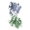
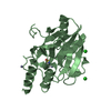
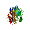
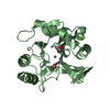
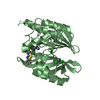
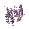
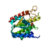
| Title | Crystal structure of the human acyl protein thioesterase 1 at 1.5 A resolution | ||||||
 Components Components | PROTEIN (ACYL PROTEIN THIOESTERASE 1) | ||||||
 Keywords Keywords |  HYDROLASE / ALPHA/BETA HYDROLASE / SERINE HYDROLASE / SAD / ANOMALOUS DIFFRACTION HYDROLASE / ALPHA/BETA HYDROLASE / SERINE HYDROLASE / SAD / ANOMALOUS DIFFRACTION | ||||||
| Function / homology |  Function and homology information Function and homology informationprotein depalmitoylation / palmitoyl[protein] hydrolase / negative regulation of Golgi to plasma membrane protein transport / palmitoyl-(protein) hydrolase activity / phospholipase activity / lipase activity / lysophospholipase activity / Hydrolases; Acting on ester bonds; Thioester hydrolases / carboxylic ester hydrolase activity / fatty acid transport ...protein depalmitoylation / palmitoyl[protein] hydrolase / negative regulation of Golgi to plasma membrane protein transport / palmitoyl-(protein) hydrolase activity / phospholipase activity / lipase activity / lysophospholipase activity / Hydrolases; Acting on ester bonds; Thioester hydrolases / carboxylic ester hydrolase activity / fatty acid transport / eNOS activation / fatty acid metabolic process / RAS processing / nuclear membrane / cell surface / endoplasmic reticulum / extracellular exosome / nucleoplasm / plasma membrane / cytosol / cytoplasmSimilarity search - Function | ||||||
| Biological species |  Homo sapiens (human) Homo sapiens (human) | ||||||
| Method | X-RAY DIFFRACTION / SYNCHROTRON / Resolution: 1.5 Å | ||||||
 Authors Authors | Devedjiev, Y. / Dauter, Z. / Kuznetsov, S. / Jones, T. / Derewenda, Z. | ||||||
 Citation Citation | Journal: Structure Fold.Des. / Year: 2000 Title: Crystal structure of the human acyl protein thioesterase I from a single X-ray data set to 1.5 A. Authors: Devedjiev, Y. / Dauter, Z. / Kuznetsov, S.R. / Jones, T.L. / Derewenda, Z.S. | ||||||
| History |
|
- Structure visualization
| Structure viewer | Molecule: MolmilJmol/JSmol |
|---|
- Downloads & links
-Download
| PDBx/mmCIF format | 1fj2.cif.gz | 110.9 KB | Display | PDBx/mmCIF format |
|---|---|---|---|---|
| PDB format | pdb1fj2.ent.gz | 83.7 KB | Display | PDB format |
| PDBx/mmJSON format | 1fj2.json.gz | Tree view | PDBx/mmJSON format | |
| Others |  Other downloads Other downloads |
-Validation report
| Arichive directory | https://data.pdbj.org/pub/pdb/validation_reports/fj/1fj2ftp://data.pdbj.org/pub/pdb/validation_reports/fj/1fj2 | HTTPS FTP |
|---|
-Related structure data
| Related structure data | |
|---|---|
| Similar structure data |


-Links
 PDBj
PDBj


- Assembly
| Deposited unit | 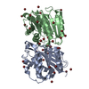
| ||||||||
|---|---|---|---|---|---|---|---|---|---|
| 1 | 
| ||||||||
| 2 | 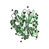
| ||||||||
| 3 |
| ||||||||
| Unit cell |
| ||||||||
| Details | IN SOLUTION, THERE IS AN EQUILLIBRIUM OF MONOMERIC AND DIMERIC SPECIES OF HUMAN ACYL PROTEIN THIOESTERASE 1. BIOLOGICAL UNIT OF THE ENZYME is STILL UNCERTAIN. |
-Components
| #1: Protein | Mass: 24920.762 Da / Num. of mol.: 2 Source method: isolated from a genetically manipulated source Source: (gene. exp.) Homo sapiens (human) / Production host:  Escherichia coli (E. coli) Escherichia coli (E. coli)References: UniProt: O75608, alkylglycerophosphoethanolamine phosphodiesterase#2: Chemical | ChemComp-BR / Bromide  Mass: 79.904 Da / Num. of mol.: 40 / Source method: obtained synthetically / Formula: Br Mass: 79.904 Da / Num. of mol.: 40 / Source method: obtained synthetically / Formula: Br#3: Water | ChemComp-HOH / | Water Mass: 18.015 Da / Num. of mol.: 465 / Source method: isolated from a natural source / Formula: H2O Mass: 18.015 Da / Num. of mol.: 465 / Source method: isolated from a natural source / Formula: H2O |
|---|
-Experimental details
-Experiment
| Experiment | Method: X-RAY DIFFRACTION / Number of used crystals: 1 |
|---|
- Sample preparation
| Crystal | Density Matthews: 1.75 Å3/Da / Density % sol: 30 % | ||||||||||||||||||||||||||||||||||||||||||||||||
|---|---|---|---|---|---|---|---|---|---|---|---|---|---|---|---|---|---|---|---|---|---|---|---|---|---|---|---|---|---|---|---|---|---|---|---|---|---|---|---|---|---|---|---|---|---|---|---|---|---|
| Crystal grow | Temperature: 293 K / Method: vapor diffusion, hanging drop / pH: 5 Details: 28 - 32% OF SATURATED AMMONIUM SULFATE, 0.1 M SODIUM ACETATE, DI-THIO-THREITOL, pH 5.0, VAPOR DIFFUSION, HANGING DROP, temperature 293K | ||||||||||||||||||||||||||||||||||||||||||||||||
| Crystal grow | *PLUS pH: 8 / Method: vapor diffusion | ||||||||||||||||||||||||||||||||||||||||||||||||
| Components of the solutions | *PLUS
|
-Data collection
| Diffraction | Mean temperature: 100 K |
|---|---|
| Diffraction source | Source: SYNCHROTRON / Site: NSLS  / Beamline: X9B / Wavelength: 0.91374 / Beamline: X9B / Wavelength: 0.91374 |
| Detector | Type: ADSC QUANTUM / Detector: CCD / Date: Apr 3, 2000 |
| Radiation | Protocol: SINGLE WAVELENGTH / Monochromatic (M) / Laue (L): M / Scattering type: x-ray |
| Radiation wavelength | Wavelength: 0.91374 Å / Relative weight: 1 |
| Reflection | Resolution: 1.48→30 Å / Num. all: 163216 / Num. obs: 50736 / % possible obs: 80 % / Observed criterion σ(F): 1 / Observed criterion σ(I): 1 / Redundancy: 2.2 % / Biso Wilson estimate: 15.2 Å2 / Rmerge(I) obs: 0.052 / Net I/σ(I): 17 |
| Reflection shell | Resolution: 1.48→1.53 Å / Redundancy: 1.6 % / Rmerge(I) obs: 0.32 / Mean I/σ(I) obs: 2.5 / % possible all: 27 |
| Reflection | *PLUS Redundancy: 3.2 % / Num. measured all: 163216 |
| Reflection shell | *PLUS % possible obs: 27.1 % / Rmerge(I) obs: 0.343 |
- Processing
| Software |
| ||||||||||||||||||||||||||||||||||||||||||||||||||||||||||||||||||||||||||||
|---|---|---|---|---|---|---|---|---|---|---|---|---|---|---|---|---|---|---|---|---|---|---|---|---|---|---|---|---|---|---|---|---|---|---|---|---|---|---|---|---|---|---|---|---|---|---|---|---|---|---|---|---|---|---|---|---|---|---|---|---|---|---|---|---|---|---|---|---|---|---|---|---|---|---|---|---|---|
| Refinement | Resolution: 1.5→20 Å / SU B: 2.2 / SU ML: 0.08 / σ(F): 1 / ESU R: 0.11 / ESU R Free: 0.12
| ||||||||||||||||||||||||||||||||||||||||||||||||||||||||||||||||||||||||||||
| Refinement step | Cycle: LAST / Resolution: 1.5→20 Å
| ||||||||||||||||||||||||||||||||||||||||||||||||||||||||||||||||||||||||||||
| Refine LS restraints |
| ||||||||||||||||||||||||||||||||||||||||||||||||||||||||||||||||||||||||||||
| Software | *PLUS Name: REFMAC / Classification: refinement | ||||||||||||||||||||||||||||||||||||||||||||||||||||||||||||||||||||||||||||
| Refinement | *PLUS Highest resolution: 1.5 Å / σ(F): 1 / % reflection Rfree: 2.5 % / Rfactor obs: 0.186 | ||||||||||||||||||||||||||||||||||||||||||||||||||||||||||||||||||||||||||||
| Solvent computation | *PLUS | ||||||||||||||||||||||||||||||||||||||||||||||||||||||||||||||||||||||||||||
| Displacement parameters | *PLUS | ||||||||||||||||||||||||||||||||||||||||||||||||||||||||||||||||||||||||||||
| Refine LS restraints | *PLUS
|
