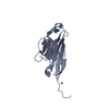
 Movie
Movie Controller


+ Open data

- Basic information
| Entry | Database: PDB / ID: 1fhg | ||||||
|---|---|---|---|---|---|---|---|
| Title | HIGH RESOLUTION REFINEMENT OF TELOKIN | ||||||
 Components Components | TELOKIN | ||||||
 Keywords Keywords |  CONTRACTILE PROTEIN / immunoglobulin fold / beta barrel CONTRACTILE PROTEIN / immunoglobulin fold / beta barrel | ||||||
| Function / homology |  Function and homology information Function and homology information | ||||||
| Biological species |  Meleagris gallopavo (turkey) Meleagris gallopavo (turkey) | ||||||
| Method | X-RAY DIFFRACTION / SYNCHROTRON / IR / Resolution: 2 Å | ||||||
 Authors Authors | Tomchick, D.R. / Minor, W. / Kiyatkin, A. / Lewinski, K. / Somlyo, A.V. / Somlyo, A.P. | ||||||
 Citation Citation | Journal: J.Mol.Biol. / Year: 1992 Title: X-ray structure determination of telokin, the C-terminal domain of myosin light chain kinase, at 2.8 A resolution. Authors: Holden, H.M. / Ito, M. / Hartshorne, D.J. / Rayment, I. #1: Journal: J.Mol.Biol. / Year: 1992Title: X-Ray Structure Determination of Telokin, the C-terminal Domain of Myosin Light Chain Kinase, at 2.8 Angstroms Resolution Authors: Holden, H.M. / Ito, M. / Hartshorne, D.J. / Rayment, I. #2: Journal: ACTA PHYSIOL.SCAND. / Year: 1998Title: Regulation of the Cross-bridge Cycle: the Effects of MgADP, LC17 Isoforms and Telokin. Authors: Somlyo, A.V. / Matthew, J.D. / Wu, X. / Khromov, A.S. / Somlyo, A.P. #3: Journal: J.Biol.Chem. / Year: 1998Title: Acceleration of Myosin Light Chain Dephosphorylation and Relaxation of Smooth Muscle by Telokin. Synergism with Cyclic Nucleotide-activated Kinase. Authors: Wu, X. / Haystead, T.A. / Nakamoto, R.K. / Somlyo, A.V. / Somlyo, A.P. | ||||||
| History |
|
- Structure visualization
| Structure viewer | Molecule: MolmilJmol/JSmol |
|---|
- Downloads & links
-Download
| PDBx/mmCIF format | 1fhg.cif.gz | 35.2 KB | Display | PDBx/mmCIF format |
|---|---|---|---|---|
| PDB format | pdb1fhg.ent.gz | 22.8 KB | Display | PDB format |
| PDBx/mmJSON format | 1fhg.json.gz | Tree view | PDBx/mmJSON format | |
| Others |  Other downloads Other downloads |
-Validation report
| Arichive directory | https://data.pdbj.org/pub/pdb/validation_reports/fh/1fhgftp://data.pdbj.org/pub/pdb/validation_reports/fh/1fhg | HTTPS FTP |
|---|
-Related structure data
| Related structure data |  1tlkSC S: Starting model for refinement C: citing same article ( |
|---|---|
| Similar structure data |


-Links
 PDBj
PDBj

- Assembly
| Deposited unit | 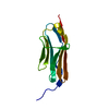
| |||||||||
|---|---|---|---|---|---|---|---|---|---|---|
| 1 |
| |||||||||
| Unit cell |
| |||||||||
| Components on special symmetry positions |
|
-Components
| #1: Protein | Mass: 16974.352 Da / Num. of mol.: 1 / Source method: isolated from a natural source / Source: (natural) Meleagris gallopavo (turkey) / Tissue: GIZZARD / References: UniProt: P56276 |
|---|---|
| #2: Water | ChemComp-HOH / Water Mass: 18.015 Da / Num. of mol.: 82 / Source method: isolated from a natural source / Formula: H2O Mass: 18.015 Da / Num. of mol.: 82 / Source method: isolated from a natural source / Formula: H2O |
-Experimental details
-Experiment
| Experiment | Method: X-RAY DIFFRACTION / Number of used crystals: 1 |
|---|
- Sample preparation
| Crystal | Density Matthews: 2.03 Å3/Da / Density % sol: 30 % |
|---|---|
| Crystal grow | Temperature: 273 K / Method: vapor diffusion, sitting drop / pH: 4 Details: PEG6000, sodium acetate, pH 4.0, VAPOR DIFFUSION, SITTING DROP, temperature 273.0K |
-Data collection
| Diffraction | Mean temperature: 100 K |
|---|---|
| Diffraction source | Source: SYNCHROTRON / Site: ESRF  / Beamline: BM14 / Wavelength: 1 / Beamline: BM14 / Wavelength: 1 |
| Detector | Detector: CCD / Date: Sep 1, 1997 |
| Radiation | Protocol: SINGLE WAVELENGTH / Monochromatic (M) / Laue (L): M / Scattering type: x-ray |
| Radiation wavelength | Wavelength: 1 Å / Relative weight: 1 |
| Reflection | Resolution: 1.98→20 Å / Num. all: 9904 / Num. obs: 9904 / % possible obs: 99.6 % / Observed criterion σ(F): 0 / Observed criterion σ(I): 0 / Redundancy: 10.5 % / Biso Wilson estimate: 47.3 Å2 / Rmerge(I) obs: 0.07 / Net I/σ(I): 35.7 |
| Reflection shell | Resolution: 1.98→2.05 Å / Redundancy: 4.7 % / Rmerge(I) obs: 0.574 / Mean I/σ(I) obs: 2.34 / Num. unique all: 967 / % possible all: 99.6 |
- Processing
| Software |
| ||||||||||||||||||||||||||||||||||||
|---|---|---|---|---|---|---|---|---|---|---|---|---|---|---|---|---|---|---|---|---|---|---|---|---|---|---|---|---|---|---|---|---|---|---|---|---|---|
| Refinement | Method to determine structure: IR Starting model: 1tlk Resolution: 2→20 Å / σ(F): 0 / σ(I): 0 / Stereochemistry target values: Engh & Huber
| ||||||||||||||||||||||||||||||||||||
| Solvent computation | Bsol: 52.72 Å2 / ksol: 0.369 e/Å3 | ||||||||||||||||||||||||||||||||||||
| Displacement parameters | Biso mean: 42.95 Å2
| ||||||||||||||||||||||||||||||||||||
| Refine analyze |
| ||||||||||||||||||||||||||||||||||||
| Refinement step | Cycle: LAST / Resolution: 2→20 Å
| ||||||||||||||||||||||||||||||||||||
| Refine LS restraints |
| ||||||||||||||||||||||||||||||||||||
| LS refinement shell | Resolution: 2→2.07 Å / Total num. of bins used: 10
| ||||||||||||||||||||||||||||||||||||
| Xplor file | Serial no: 1 / Param file: protein_rep.param / Topol file: protein.top |
