 Movie
Movie Controller


+ Open data

- Basic information
| Entry | Database: PDB / ID: 1fdr | ||||||
|---|---|---|---|---|---|---|---|
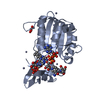
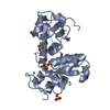
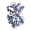
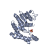
| Title | FLAVODOXIN REDUCTASE FROM E. COLI | ||||||
 Components Components | FLAVODOXIN REDUCTASE | ||||||
 Keywords Keywords |  FLAVOPROTEIN / FLAVODOXIN REDUCTASE / FERREDOXIN REDUCTASE / FLAVIN / OXIDOREDUCTASE FLAVOPROTEIN / FLAVODOXIN REDUCTASE / FERREDOXIN REDUCTASE / FLAVIN / OXIDOREDUCTASE | ||||||
| Function / homology |  Function and homology information Function and homology informationflavodoxin-NADP+ reductase / aconitate hydratase activity / succinate dehydrogenase activity / ferredoxin-NADP+ reductase / ferredoxin-NADP+ reductase activity / heme catabolic process / response to superoxide / iron-sulfur cluster assembly / FAD binding / cellular response to oxidative stress ...flavodoxin-NADP+ reductase / aconitate hydratase activity / succinate dehydrogenase activity / ferredoxin-NADP+ reductase / ferredoxin-NADP+ reductase activity / heme catabolic process / response to superoxide / iron-sulfur cluster assembly / FAD binding / cellular response to oxidative stress / response to xenobiotic stimulus / cytosolSimilarity search - Function | ||||||
| Biological species |  Escherichia coli (E. coli) Escherichia coli (E. coli) | ||||||
| Method | X-RAY DIFFRACTION / MIR / Resolution: 1.7 Å | ||||||
 Authors Authors | Ingelman, M. / Bianchi, V. / Eklund, H. | ||||||
 Citation Citation | Journal: J.Mol.Biol. / Year: 1997 Title: The three-dimensional structure of flavodoxin reductase from Escherichia coli at 1.7 A resolution. Authors: Ingelman, M. / Bianchi, V. / Eklund, H. | ||||||
| History |
|
- Structure visualization
| Structure viewer | Molecule: MolmilJmol/JSmol |
|---|
- Downloads & links
-Download
| PDBx/mmCIF format | 1fdr.cif.gz | 65.7 KB | Display | PDBx/mmCIF format |
|---|---|---|---|---|
| PDB format | pdb1fdr.ent.gz | 47.9 KB | Display | PDB format |
| PDBx/mmJSON format | 1fdr.json.gz | Tree view | PDBx/mmJSON format | |
| Others |  Other downloads Other downloads |
-Validation report
| Arichive directory | https://data.pdbj.org/pub/pdb/validation_reports/fd/1fdrftp://data.pdbj.org/pub/pdb/validation_reports/fd/1fdr | HTTPS FTP |
|---|
-Related structure data
| Similar structure data |
|---|


-Links
 PDBj
PDBj

- Assembly
| Deposited unit | 
| ||||||||
|---|---|---|---|---|---|---|---|---|---|
| 1 |
| ||||||||
| Unit cell |
|
-Components
| #1: Protein | Mass: 27812.979 Da / Num. of mol.: 1 / Mutation: R126Q Source method: isolated from a genetically manipulated source Source: (gene. exp.) Escherichia coli (E. coli) / Strain: K12 C600 / Cellular location: CYTOPLASM / Gene: FPR / Plasmid: PEE1010 / Cellular location (production host): CYTOPLASM / Production host: Escherichia coli (E. coli) / Strain (production host): S30 / References: UniProt: P28861, ferredoxin-NADP+ reductase |
|---|---|
| #2: Chemical | ChemComp-FAD / Flavin adenine dinucleotide  Mass: 785.550 Da / Num. of mol.: 1 / Source method: obtained synthetically / Formula: C27H33N9O15P2 / Comment: FAD*YM Mass: 785.550 Da / Num. of mol.: 1 / Source method: obtained synthetically / Formula: C27H33N9O15P2 / Comment: FAD*YM |
| #3: Water | ChemComp-HOH / Water Mass: 18.015 Da / Num. of mol.: 205 / Source method: isolated from a natural source / Formula: H2O Mass: 18.015 Da / Num. of mol.: 205 / Source method: isolated from a natural source / Formula: H2O |
-Experimental details
-Experiment
| Experiment | Method: X-RAY DIFFRACTION / Number of used crystals: 1 |
|---|
- Sample preparation
| Crystal | Density Matthews: 2.3 Å3/Da / Density % sol: 47 % | ||||||||||||||||||||
|---|---|---|---|---|---|---|---|---|---|---|---|---|---|---|---|---|---|---|---|---|---|
| Crystal grow | pH: 7.3 / Details: 1.2 M SODIUM CITRATE 100MM TRISHCL PH 7.3 | ||||||||||||||||||||
| Crystal grow | *PLUS Temperature: 20 ℃ / Method: vapor diffusion | ||||||||||||||||||||
| Components of the solutions | *PLUS
|
-Data collection
| Diffraction | Mean temperature: 277 K |
|---|---|
| Diffraction source | Source: ROTATING ANODE / Type: RIGAKU RUH2R / Wavelength: 1.5418 |
| Detector | Type: RIGAKU / Detector: IMAGE PLATE / Date: Mar 8, 1994 / Details: MIRRORS |
| Radiation | Monochromator: NI FILTER / Monochromatic (M) / Laue (L): M / Scattering type: x-ray |
| Radiation wavelength | Wavelength: 1.5418 Å / Relative weight: 1 |
| Reflection | Resolution: 1.7→20 Å / Num. obs: 28503 / % possible obs: 96 % / Observed criterion σ(I): 2 / Redundancy: 2.5 % / Rsym value: 0.064 / Net I/σ(I): 17.8 |
| Reflection shell | Resolution: 1.7→2 Å / Redundancy: 1 % / Mean I/σ(I) obs: 3.3 / Rsym value: 0.23 / % possible all: 29 |
| Reflection | *PLUS Num. measured all: 68969 / Rmerge(I) obs: 0.064 |
| Reflection shell | *PLUS % possible obs: 29 % / Rmerge(I) obs: 0.23 |
- Processing
| Software |
| ||||||||||||||||||||||||||||||||||||||||||||||||||||||||||||
|---|---|---|---|---|---|---|---|---|---|---|---|---|---|---|---|---|---|---|---|---|---|---|---|---|---|---|---|---|---|---|---|---|---|---|---|---|---|---|---|---|---|---|---|---|---|---|---|---|---|---|---|---|---|---|---|---|---|---|---|---|---|
| Refinement | Method to determine structure: MIR / Resolution: 1.7→8 Å / Data cutoff high absF: 1000000 / Data cutoff low absF: 0.1 / σ(F): 2
| ||||||||||||||||||||||||||||||||||||||||||||||||||||||||||||
| Displacement parameters | Biso mean: 18.1 Å2 | ||||||||||||||||||||||||||||||||||||||||||||||||||||||||||||
| Refine analyze | Luzzati coordinate error obs: 0.21 Å / Luzzati d res low obs: 10 Å | ||||||||||||||||||||||||||||||||||||||||||||||||||||||||||||
| Refinement step | Cycle: LAST / Resolution: 1.7→8 Å
| ||||||||||||||||||||||||||||||||||||||||||||||||||||||||||||
| Refine LS restraints |
| ||||||||||||||||||||||||||||||||||||||||||||||||||||||||||||
| LS refinement shell | Resolution: 1.7→1.87 Å / Total num. of bins used: 6
| ||||||||||||||||||||||||||||||||||||||||||||||||||||||||||||
| Xplor file |
| ||||||||||||||||||||||||||||||||||||||||||||||||||||||||||||
| Software | *PLUS Name: X-PLOR / Version: 3.1 / Classification: refinement | ||||||||||||||||||||||||||||||||||||||||||||||||||||||||||||
| Refinement | *PLUS | ||||||||||||||||||||||||||||||||||||||||||||||||||||||||||||
| Solvent computation | *PLUS | ||||||||||||||||||||||||||||||||||||||||||||||||||||||||||||
| Displacement parameters | *PLUS | ||||||||||||||||||||||||||||||||||||||||||||||||||||||||||||
| Refine LS restraints | *PLUS
| ||||||||||||||||||||||||||||||||||||||||||||||||||||||||||||
| LS refinement shell | *PLUS Rfactor obs: 0.29 |
