 Movie
Movie Controller

[English] 日本語
 Yorodumi
- PDB-1f6v: SOLUTION STRUCTURE OF THE C TERMINAL OF MU B TRANSPOSITION PROTEIN -
+ Open data

- Basic information
| Entry | Database: PDB / ID: 1f6v | ||||||
|---|---|---|---|---|---|---|---|
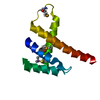
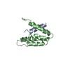
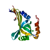
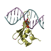
| Title | SOLUTION STRUCTURE OF THE C TERMINAL OF MU B TRANSPOSITION PROTEIN | ||||||
 Components Components | DNA TRANSPOSITION PROTEIN | ||||||
 Keywords Keywords |  DNA BINDING PROTEIN / Mu phage / recombination / transposition / ATPase / DNA binding / high salt / solution structure DNA BINDING PROTEIN / Mu phage / recombination / transposition / ATPase / DNA binding / high salt / solution structure | ||||||
| Function / homology |  Function and homology information Function and homology informationDNA transposition / viral DNA genome replication / Hydrolases; Acting on acid anhydrides; In phosphorus-containing anhydrides / DNA integration / DNA replication / host cell cytoplasm / ATP hydrolysis activity / DNA binding / ATP binding / metal ion bindingSimilarity search - Function | ||||||
| Biological species |  Enterobacteria phage Mu (virus) Enterobacteria phage Mu (virus) | ||||||
| Method | SOLUTION NMR / simulated annealing | ||||||
 Authors Authors | Hung, L.-H. / Chaconas, G. / Shaw, G.S. | ||||||
 Citation Citation | Journal: EMBO J. / Year: 2000 Title: The solution structure of the C-terminal domain of the Mu B transposition protein. Authors: Hung, L.H. / Chaconas, G. / Shaw, G.S. | ||||||
| History |
|
- Structure visualization
| Structure viewer | Molecule: MolmilJmol/JSmol |
|---|
- Downloads & links
-Download
| PDBx/mmCIF format | 1f6v.cif.gz | 575 KB | Display | PDBx/mmCIF format |
|---|---|---|---|---|
| PDB format | pdb1f6v.ent.gz | 477.5 KB | Display | PDB format |
| PDBx/mmJSON format | 1f6v.json.gz | Tree view | PDBx/mmJSON format | |
| Others |  Other downloads Other downloads |
-Validation report
| Arichive directory | https://data.pdbj.org/pub/pdb/validation_reports/f6/1f6vftp://data.pdbj.org/pub/pdb/validation_reports/f6/1f6v | HTTPS FTP |
|---|
-Related structure data
| Similar structure data |
|---|


-Links
 PDBj
PDBj


- Assembly
| Deposited unit | 
| |||||||||
|---|---|---|---|---|---|---|---|---|---|---|
| 1 |
| |||||||||
| NMR ensembles |
|
-Components
| #1: Protein | Mass: 10237.761 Da / Num. of mol.: 1 / Fragment: C-TERMINAL DOMAIN Source method: isolated from a genetically manipulated source Source: (gene. exp.) Enterobacteria phage Mu (virus) / Genus: Mu-like viruses / Description: T7 PHAGE / Plasmid: PHH05 / Production host:  Escherichia coli (E. coli) / References: UniProt: P03763 Escherichia coli (E. coli) / References: UniProt: P03763 |
|---|
-Experimental details
-Experiment
| Experiment | Method: SOLUTION NMR | ||||||||||||||||||||||||||||||||
|---|---|---|---|---|---|---|---|---|---|---|---|---|---|---|---|---|---|---|---|---|---|---|---|---|---|---|---|---|---|---|---|---|---|
| NMR experiment |
| ||||||||||||||||||||||||||||||||
| NMR details | Text: High ionic conditions precluded many of the standard triple resonance experiments. |
- Sample preparation
| Details |
| |||||||||||||||
|---|---|---|---|---|---|---|---|---|---|---|---|---|---|---|---|---|
| Sample conditions | Ionic strength: 1.5 M NaCl / pH: 6.8 / Pressure: ambient / Temperature: 298 K | |||||||||||||||
| Crystal grow | *PLUS Method: other / Details: NMR |
-NMR measurement
| NMR spectrometer | Type: Varian UNITY / Manufacturer: Varian / Model: UNITY / Field strength: 500 MHz |
|---|
- Processing
| NMR software |
| ||||||||||||||||
|---|---|---|---|---|---|---|---|---|---|---|---|---|---|---|---|---|---|
| Refinement | Method: simulated annealing / Software ordinal: 1 Details: The structures are based on a total of 1047 NOE derived, and 55 dihedral constraints. There are no long-range NOEs observed for the first 8 residues which are disordered. | ||||||||||||||||
| NMR representative | Selection criteria: lowest energy | ||||||||||||||||
| NMR ensemble | Conformer selection criteria: structures with lowest energy and no restraint violations with phi psi angles in allowed regions Conformers calculated total number: 100 / Conformers submitted total number: 20 |
