 Movie
Movie Controller


+ Open data

- Basic information
| Entry | Database: PDB / ID: 1f4b | ||||||
|---|---|---|---|---|---|---|---|
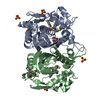
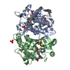
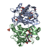
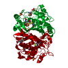
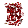
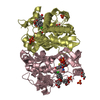
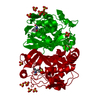
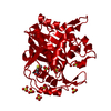
| Title | CRYSTAL STRUCTURE OF ESCHERICHIA COLI THYMIDYLATE SYNTHASE | ||||||
 Components Components | THYMIDYLATE SYNTHASE | ||||||
 Keywords Keywords | TRANSFERASE / E. coli thymidylate synthase | ||||||
| Function / homology |  Function and homology informationthymidylate synthase / thymidylate synthase activity / dTMP biosynthetic process / dTTP biosynthetic process / response to radiation / regulation of translation / methylation / magnesium ion binding / protein homodimerization activity / RNA binding / cytosol Function and homology informationthymidylate synthase / thymidylate synthase activity / dTMP biosynthetic process / dTTP biosynthetic process / response to radiation / regulation of translation / methylation / magnesium ion binding / protein homodimerization activity / RNA binding / cytosolSimilarity search - Function | ||||||
| Biological species |  Escherichia coli (E. coli) Escherichia coli (E. coli) | ||||||
| Method | X-RAY DIFFRACTION / Resolution: 1.75 Å | ||||||
 Authors Authors | Erlanson, D.A. / Braisted, A.C. / Raphael, D.R. / Randal, M. / Stroud, R.M. / Gordon, E. / Wells, J.A. | ||||||
 Citation Citation | Journal: Proc.Natl.Acad.Sci.USA / Year: 2000 Title: Site-directed ligand discovery. Authors: Erlanson, D.A. / Braisted, A.C. / Raphael, D.R. / Randal, M. / Stroud, R.M. / Gordon, E.M. / Wells, J.A. | ||||||
| History |
|
- Structure visualization
| Structure viewer | Molecule: MolmilJmol/JSmol |
|---|
- Downloads & links
-Download
| PDBx/mmCIF format | 1f4b.cif.gz | 68.7 KB | Display | PDBx/mmCIF format |
|---|---|---|---|---|
| PDB format | pdb1f4b.ent.gz | 55.3 KB | Display | PDB format |
| PDBx/mmJSON format | 1f4b.json.gz | Tree view | PDBx/mmJSON format | |
| Others |  Other downloads Other downloads |
-Validation report
| Arichive directory | https://data.pdbj.org/pub/pdb/validation_reports/f4/1f4bftp://data.pdbj.org/pub/pdb/validation_reports/f4/1f4b | HTTPS FTP |
|---|
-Related structure data


-Links
 PDBj
PDBj- Assembly
| Deposited unit | 
| |||||||||
|---|---|---|---|---|---|---|---|---|---|---|
| 1 | 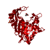
| |||||||||
| Unit cell |
| |||||||||
| Components on special symmetry positions |
| |||||||||
| Details | The biological assembly is a dimer constructed from chain A and a symmetry partner generated by the two-fold |
-Components
| #1: Protein | Mass: 30559.666 Da / Num. of mol.: 1 Source method: isolated from a genetically manipulated source Source: (gene. exp.) Escherichia coli (E. coli) / Plasmid: PTHYAWT / Production host: Escherichia coli (E. coli) / Strain (production host): C2913 / References: UniProt: P0A884, thymidylate synthase | ||||
|---|---|---|---|---|---|
| #2: Chemical | Sulfate  Mass: 96.063 Da / Num. of mol.: 3 / Source method: obtained synthetically / Formula: SO4 Mass: 96.063 Da / Num. of mol.: 3 / Source method: obtained synthetically / Formula: SO4#3: Chemical | Glycerol  Mass: 92.094 Da / Num. of mol.: 2 / Source method: obtained synthetically / Formula: C3H8O3 Mass: 92.094 Da / Num. of mol.: 2 / Source method: obtained synthetically / Formula: C3H8O3#4: Water | ChemComp-HOH / | Water Mass: 18.015 Da / Num. of mol.: 262 / Source method: isolated from a natural source / Formula: H2O Mass: 18.015 Da / Num. of mol.: 262 / Source method: isolated from a natural source / Formula: H2O |
-Experimental details
-Experiment
| Experiment | Method: X-RAY DIFFRACTION / Number of used crystals: 1 |
|---|
- Sample preparation
| Crystal | Density Matthews: 3.09 Å3/Da / Density % sol: 60 % | ||||||||||||||||||||||||||||||||||||||||||||||||||
|---|---|---|---|---|---|---|---|---|---|---|---|---|---|---|---|---|---|---|---|---|---|---|---|---|---|---|---|---|---|---|---|---|---|---|---|---|---|---|---|---|---|---|---|---|---|---|---|---|---|---|---|
| Crystal grow | Temperature: 293 K / Method: vapor diffusion, hanging drop / pH: 7 Details: 2.0 M ammonium sulphate, 20 mM potassium phosphate, 0.2 M EDTA, pH 7.0, VAPOR DIFFUSION, HANGING DROP | ||||||||||||||||||||||||||||||||||||||||||||||||||
| Crystal grow | *PLUS Method: vapor diffusionDetails: Perry, K.M., (1990) Proteins: Struct.,Funct., Genet., 8, 315. | ||||||||||||||||||||||||||||||||||||||||||||||||||
| Components of the solutions | *PLUS
|
-Data collection
| Diffraction | Mean temperature: 100 K |
|---|---|
| Diffraction source | Source: ROTATING ANODE / Type: RIGAKU RUH3R / Wavelength: 1.5418 |
| Detector | Type: RIGAKU RAXIS IV / Detector: IMAGE PLATE / Date: Dec 5, 1999 |
| Radiation | Protocol: SINGLE WAVELENGTH / Monochromatic (M) / Laue (L): M / Scattering type: x-ray |
| Radiation wavelength | Wavelength: 1.5418 Å / Relative weight: 1 |
| Reflection | Resolution: 1.75→10 Å / Num. all: 36586 / Num. obs: 36586 / % possible obs: 96.7 % / Observed criterion σ(F): 0 / Observed criterion σ(I): 0 / Redundancy: 2.8 % / Biso Wilson estimate: 31.9 Å2 / Rmerge(I) obs: 0.049 / Net I/σ(I): 20.5 |
| Reflection shell | Resolution: 1.75→1.81 Å / Redundancy: 2 % / Rmerge(I) obs: 0.338 / Mean I/σ(I) obs: 4 / Num. unique all: 3443 / % possible all: 91.6 |
| Reflection | *PLUS Num. measured all: 104019 |
| Reflection shell | *PLUS % possible obs: 91.6 % |
- Processing
| Software |
| ||||||||||||||||||||||||||||||||||||||||||||||||||||||||||||||||||||
|---|---|---|---|---|---|---|---|---|---|---|---|---|---|---|---|---|---|---|---|---|---|---|---|---|---|---|---|---|---|---|---|---|---|---|---|---|---|---|---|---|---|---|---|---|---|---|---|---|---|---|---|---|---|---|---|---|---|---|---|---|---|---|---|---|---|---|---|---|---|
| Refinement | Resolution: 1.75→10 Å / SU B: 2.84 / SU ML: 0.089 / Cross valid method: THROUGHOUT / σ(F): 0 / σ(I): 0 / ESU R: 0.11 / ESU R Free: 0.12 / Stereochemistry target values: Engh & Huber
| ||||||||||||||||||||||||||||||||||||||||||||||||||||||||||||||||||||
| Displacement parameters | Biso mean: 37.15 Å2 | ||||||||||||||||||||||||||||||||||||||||||||||||||||||||||||||||||||
| Refinement step | Cycle: LAST / Resolution: 1.75→10 Å
| ||||||||||||||||||||||||||||||||||||||||||||||||||||||||||||||||||||
| Refine LS restraints |
| ||||||||||||||||||||||||||||||||||||||||||||||||||||||||||||||||||||
| Software | *PLUS Name: REFMAC / Classification: refinement | ||||||||||||||||||||||||||||||||||||||||||||||||||||||||||||||||||||
| Refinement | *PLUS σ(F): 0 / Rfactor all: 0.2 / Rfactor obs: 0.198 | ||||||||||||||||||||||||||||||||||||||||||||||||||||||||||||||||||||
| Solvent computation | *PLUS | ||||||||||||||||||||||||||||||||||||||||||||||||||||||||||||||||||||
| Displacement parameters | *PLUS | ||||||||||||||||||||||||||||||||||||||||||||||||||||||||||||||||||||
| Refine LS restraints | *PLUS
|
