 Movie
Movie Controller

[English] 日本語
 Yorodumi
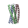
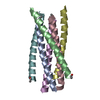
- PDB-1f23: CONTRIBUTION OF A BURIED HYDROGEN BOND TO HIV-1 ENVELOPE GLYCOPRO... -
+ Open data

- Basic information
| Entry | Database: PDB / ID: 1f23 | ||||||
|---|---|---|---|---|---|---|---|
| Title | CONTRIBUTION OF A BURIED HYDROGEN BOND TO HIV-1 ENVELOPE GLYCOPROTEIN STRUCTURE AND FUNCTION | ||||||
 Components Components | TRANSMEMBRANE GLYCOPROTEIN | ||||||
 Keywords Keywords |  VIRAL PROTEIN / HIV-1 envelope protein / gp41 / membrane fusion / HIV-1 entry VIRAL PROTEIN / HIV-1 envelope protein / gp41 / membrane fusion / HIV-1 entry | ||||||
| Function / homology |  Function and homology information Function and homology informationvirus-mediated perturbation of host defense response => GO:0019049 / : / positive regulation of plasma membrane raft polarization / positive regulation of receptor clustering / positive regulation of establishment of T cell polarity / host cell endosome membrane / clathrin-dependent endocytosis of virus by host cell / membrane => GO:0016020 / viral protein processing / fusion of virus membrane with host plasma membrane ...virus-mediated perturbation of host defense response => GO:0019049 / : / positive regulation of plasma membrane raft polarization / positive regulation of receptor clustering / positive regulation of establishment of T cell polarity / host cell endosome membrane / clathrin-dependent endocytosis of virus by host cell / membrane => GO:0016020 / viral protein processing / fusion of virus membrane with host plasma membrane / fusion of virus membrane with host endosome membrane / viral envelope / virion attachment to host cell / host cell plasma membrane / virion membrane / structural molecule activity / plasma membraneSimilarity search - Function | ||||||
| Biological species |   Human immunodeficiency virus 1 Human immunodeficiency virus 1 | ||||||
| Method | X-RAY DIFFRACTION / Resolution: 2.3 Å | ||||||
 Authors Authors | Liu, J. / Shu, W. / Fagan, M. / Nunberg, J.H. / Lu, M. | ||||||
 Citation Citation | Journal: Biochemistry / Year: 2001 Title: Structural and functional analysis of the HIV gp41 core containing an Ile573 to Thr substitution: implications for membrane fusion. Authors: Liu, J. / Shu, W. / Fagan, M.B. / Nunberg, J.H. / Lu, M. | ||||||
| History |
|
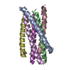
- Structure visualization
| Structure viewer | Molecule: MolmilJmol/JSmol |
|---|
- Downloads & links
-Download
| PDBx/mmCIF format | 1f23.cif.gz | 99.6 KB | Display | PDBx/mmCIF format |
|---|---|---|---|---|
| PDB format | pdb1f23.ent.gz | 78.1 KB | Display | PDB format |
| PDBx/mmJSON format | 1f23.json.gz | Tree view | PDBx/mmJSON format | |
| Others |  Other downloads Other downloads |
-Validation report
| Arichive directory | https://data.pdbj.org/pub/pdb/validation_reports/f2/1f23ftp://data.pdbj.org/pub/pdb/validation_reports/f2/1f23 | HTTPS FTP |
|---|
-Related structure data
| Similar structure data |
|---|


-Links
 PDBj
PDBj


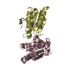
- Assembly
| Deposited unit | 
| ||||||||
|---|---|---|---|---|---|---|---|---|---|
| 1 | 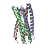
| ||||||||
| 2 | 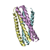
| ||||||||
| Unit cell |
|
-Components
| #1: Protein | Mass: 8820.789 Da / Num. of mol.: 6 / Fragment: ECTODOMAIN / Mutation: I28T Source method: isolated from a genetically manipulated source Source: (gene. exp.) Human immunodeficiency virus 1 / Genus: Lentivirus / Plasmid: PN36(L)C34 / Production host:  Escherichia coli (E. coli) / References: UniProt: Q89797 Escherichia coli (E. coli) / References: UniProt: Q89797#2: Water | ChemComp-HOH / | Water Mass: 18.015 Da / Num. of mol.: 271 / Source method: isolated from a natural source / Formula: H2O Mass: 18.015 Da / Num. of mol.: 271 / Source method: isolated from a natural source / Formula: H2O |
|---|
-Experimental details
-Experiment
| Experiment | Method: X-RAY DIFFRACTION / Number of used crystals: 1 |
|---|
- Sample preparation
| Crystal grow | Temperature: 293 K / Method: vapor diffusion, hanging drop / pH: 4.6 Details: Sodium citrate, PEG 4000, propanol, pH 4.6, VAPOR DIFFUSION, HANGING DROP, temperature 293K | |||||||||||||||||||||||||
|---|---|---|---|---|---|---|---|---|---|---|---|---|---|---|---|---|---|---|---|---|---|---|---|---|---|---|
| Crystal grow | *PLUS | |||||||||||||||||||||||||
| Components of the solutions | *PLUS
|
-Data collection
| Diffraction | Mean temperature: 95 K |
|---|---|
| Diffraction source | Source: ROTATING ANODE / Type: RIGAKU RU200 / Wavelength: 1.5418 |
| Detector | Type: RIGAKU RAXIS IV / Detector: IMAGE PLATE / Date: Sep 24, 1999 |
| Radiation | Protocol: SINGLE WAVELENGTH / Monochromatic (M) / Laue (L): M / Scattering type: x-ray |
| Radiation wavelength | Wavelength: 1.5418 Å / Relative weight: 1 |
| Reflection | Resolution: 2.3→50 Å / Num. all: 14667 / Num. obs: 14667 / % possible obs: 96.1 % / Observed criterion σ(F): 0 / Observed criterion σ(I): 0 / Redundancy: 2 % / Biso Wilson estimate: 20.7 Å2 / Rmerge(I) obs: 0.076 / Net I/σ(I): 7.7 |
| Reflection shell | Resolution: 2.3→2.38 Å / Redundancy: 1.9 % / Rmerge(I) obs: 0.287 / Num. unique all: 1399 / % possible all: 92.8 |
| Reflection | *PLUS Num. measured all: 62249 |
- Processing
| Software |
| ||||||||||||||||||||||||||||||||||||||||
|---|---|---|---|---|---|---|---|---|---|---|---|---|---|---|---|---|---|---|---|---|---|---|---|---|---|---|---|---|---|---|---|---|---|---|---|---|---|---|---|---|---|
| Refinement | Resolution: 2.3→50 Å / Rfactor Rfree error: 0.007 / Data cutoff high absF: 730014.41 / Data cutoff low absF: 0 / Isotropic thermal model: RESTRAINED / Cross valid method: THROUGHOUT / σ(F): 0 / σ(I): 0 / Stereochemistry target values: Engh & Huber
| ||||||||||||||||||||||||||||||||||||||||
| Solvent computation | Solvent model: FLAT MODEL / Bsol: 34.82 Å2 / ksol: 0.294 e/Å3 | ||||||||||||||||||||||||||||||||||||||||
| Displacement parameters | Biso mean: 28.5 Å2
| ||||||||||||||||||||||||||||||||||||||||
| Refine analyze |
| ||||||||||||||||||||||||||||||||||||||||
| Refinement step | Cycle: LAST / Resolution: 2.3→50 Å
| ||||||||||||||||||||||||||||||||||||||||
| Refine LS restraints |
| ||||||||||||||||||||||||||||||||||||||||
| LS refinement shell | Resolution: 2.3→2.44 Å / Rfactor Rfree error: 0.023 / Total num. of bins used: 6
| ||||||||||||||||||||||||||||||||||||||||
| Xplor file |
| ||||||||||||||||||||||||||||||||||||||||
| Software | *PLUS Name: CNS / Version: 0.5 / Classification: refinement | ||||||||||||||||||||||||||||||||||||||||
| Refinement | *PLUS σ(F): 0 / % reflection Rfree: 10.2 % / Rfactor obs: 0.191 / Rfactor Rfree: 0.27 | ||||||||||||||||||||||||||||||||||||||||
| Solvent computation | *PLUS | ||||||||||||||||||||||||||||||||||||||||
| Displacement parameters | *PLUS Biso mean: 28.5 Å2 | ||||||||||||||||||||||||||||||||||||||||
| Refine LS restraints | *PLUS
| ||||||||||||||||||||||||||||||||||||||||
| LS refinement shell | *PLUS Rfactor Rfree: 0.35 / % reflection Rfree: 10.8 % / Rfactor Rwork: 0.239 |
