 Movie
Movie Controller


+ Open data

- Basic information
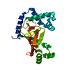
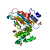
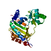
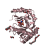
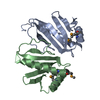
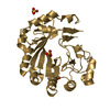
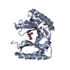
| Entry | Database: PDB / ID: 1eyv | ||||||
|---|---|---|---|---|---|---|---|
| Title | THE CRYSTAL STRUCTURE OF NUSB FROM MYCOBACTERIUM TUBERCULOSIS | ||||||
 Components Components | N-UTILIZING SUBSTANCE PROTEIN B HOMOLOG | ||||||
 Keywords Keywords |  TRANSCRIPTION / helical bundle / Structural Genomics / PSI / Protein Structure Initiative / TB Structural Genomics Consortium / TBSGC TRANSCRIPTION / helical bundle / Structural Genomics / PSI / Protein Structure Initiative / TB Structural Genomics Consortium / TBSGC | ||||||
| Function / homology |  Function and homology information Function and homology informationtranscription antitermination / DNA-templated transcription termination / RNA binding / cytosolSimilarity search - Function | ||||||
| Biological species |   Mycobacterium tuberculosis (bacteria) Mycobacterium tuberculosis (bacteria) | ||||||
| Method | X-RAY DIFFRACTION / SYNCHROTRON / Resolution: 1.6 Å | ||||||
 Authors Authors | Gopal, B. / Haire, L.F. / Cox, R.A. / Colston, M.J. / Major, S. / Brannigan, J.A. / Smerdon, S.J. / Dodson, G.G. / TB Structural Genomics Consortium (TBSGC) | ||||||
 Citation Citation | Journal: Nat.Struct.Biol. / Year: 2000 Title: The crystal structure of NusB from Mycobacterium tuberculosis. Authors: Gopal, B. / Haire, L.F. / Cox, R.A. / Colston, M.J. / Major, S. / Brannigan, J.A. / Smerdon, S.J. / Dodson, G. #1: Journal: Acta Crystallogr.,Sect.D / Year: 2000Title: Crystallization and preliminary X-ray diffraction studies on the N-utilizing substance-B (NusB) from Mycobacterium tuberculosis. Authors: Gopal, B. / Cox, R.A. / Colston, M.J. / Dodson, G.G. / Smerdon, S.J. / Haire, L.F. | ||||||
| History |
|
- Structure visualization
| Structure viewer | Molecule: MolmilJmol/JSmol |
|---|
- Downloads & links
-Download
| PDBx/mmCIF format | 1eyv.cif.gz | 67.8 KB | Display | PDBx/mmCIF format |
|---|---|---|---|---|
| PDB format | pdb1eyv.ent.gz | 51.1 KB | Display | PDB format |
| PDBx/mmJSON format | 1eyv.json.gz | Tree view | PDBx/mmJSON format | |
| Others |  Other downloads Other downloads |
-Validation report
| Arichive directory | https://data.pdbj.org/pub/pdb/validation_reports/ey/1eyvftp://data.pdbj.org/pub/pdb/validation_reports/ey/1eyv | HTTPS FTP |
|---|
-Related structure data
| Similar structure data | |
|---|---|
| Other databases |


-Links
 PDBj
PDBj- Assembly
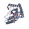
| Deposited unit | 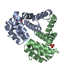
| ||||||||
|---|---|---|---|---|---|---|---|---|---|
| 1 |
| ||||||||
| Unit cell |
| ||||||||
| Details | The biological assembly is a dimer constructed from chain A a symmetry partner generated by the two-fold. |
-Components
| #1: Protein | Mass: 16765.174 Da / Num. of mol.: 2 Source method: isolated from a genetically manipulated source Source: (gene. exp.) Mycobacterium tuberculosis (bacteria) / Plasmid: PET-15B / Production host: Escherichia coli (E. coli) / References: UniProt: P95020, UniProt: P9WIV1*PLUS#2: Chemical | Phosphate  Mass: 94.971 Da / Num. of mol.: 2 / Source method: obtained synthetically / Formula: PO4 Mass: 94.971 Da / Num. of mol.: 2 / Source method: obtained synthetically / Formula: PO4#3: Water | ChemComp-HOH / | Water Mass: 18.015 Da / Num. of mol.: 291 / Source method: isolated from a natural source / Formula: H2O Mass: 18.015 Da / Num. of mol.: 291 / Source method: isolated from a natural source / Formula: H2O |
|---|
-Experimental details
-Experiment
| Experiment | Method: X-RAY DIFFRACTION / Number of used crystals: 2 |
|---|
- Sample preparation
| Crystal | Density Matthews: 1.98 Å3/Da / Density % sol: 37.75 % | |||||||||||||||||||||||||
|---|---|---|---|---|---|---|---|---|---|---|---|---|---|---|---|---|---|---|---|---|---|---|---|---|---|---|
| Crystal grow | Temperature: 295 K / Method: vapor diffusion, hanging drop / pH: 7.5 Details: 2.0 M ammonium sulfate, 0.1 M Na HEPES buffer, pH 7.5, 2 % PEG 400, VAPOR DIFFUSION, HANGING DROP, temperature 22K | |||||||||||||||||||||||||
| Crystal grow | *PLUS Temperature: 291 KDetails: Gopal, B., (2000) Acta Crystallogr., Sect.D, 56, 64. | |||||||||||||||||||||||||
| Components of the solutions | *PLUS
|
-Data collection
| Diffraction |
| |||||||||||||||
|---|---|---|---|---|---|---|---|---|---|---|---|---|---|---|---|---|
| Diffraction source |
| |||||||||||||||
| Detector | Date: May 26, 1999 | |||||||||||||||
| Radiation | Protocol: SINGLE WAVELENGTH / Monochromatic (M) / Laue (L): M / Scattering type: x-ray | |||||||||||||||
| Radiation wavelength | Wavelength: 0.87 Å / Relative weight: 1 | |||||||||||||||
| Reflection | Resolution: 1.6→15 Å / Num. all: 1501196 / Num. obs: 34118 / % possible obs: 95.5 % / Observed criterion σ(F): 2 / Redundancy: 4.4 % / Biso Wilson estimate: 16.7 Å2 / Rmerge(I) obs: 0.052 | |||||||||||||||
| Reflection shell | Resolution: 1.6→1.63 Å / Redundancy: 4.3 % / Rmerge(I) obs: 0.331 / Num. unique all: 1328 / % possible all: 75 | |||||||||||||||
| Reflection shell | *PLUS % possible obs: 75 % |


- Processing
| Software |
| |||||||||||||||||||||||||
|---|---|---|---|---|---|---|---|---|---|---|---|---|---|---|---|---|---|---|---|---|---|---|---|---|---|---|
| Refinement | Resolution: 1.6→15 Å / σ(F): 2
| |||||||||||||||||||||||||
| Refinement step | Cycle: LAST / Resolution: 1.6→15 Å
| |||||||||||||||||||||||||
| Refine LS restraints |
| |||||||||||||||||||||||||
| Software | *PLUS Name: REFMAC / Classification: refinement | |||||||||||||||||||||||||
| Refine LS restraints | *PLUS
|
