 Movie
Movie Controller


+ Open data

- Basic information
| Entry | Database: PDB / ID: 1es6 | ||||||
|---|---|---|---|---|---|---|---|
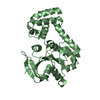
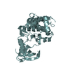
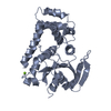
| Title | CRYSTAL STRUCTURE OF THE MATRIX PROTEIN OF EBOLA VIRUS | ||||||
 Components Components | MATRIX PROTEIN VP40 | ||||||
 Keywords Keywords |  VIRAL PROTEIN / beta sandwich / anti-parallel strands / beta sheet / helix VIRAL PROTEIN / beta sandwich / anti-parallel strands / beta sheet / helix | ||||||
| Function / homology |  Function and homology information Function and homology informationhost cell endomembrane system / host cell late endosome membrane / viral budding via host ESCRT complex / structural constituent of virion / ribonucleoprotein complex / host cell plasma membrane / virion membrane / RNA binding / extracellular region / membraneSimilarity search - Function | ||||||
| Biological species |  Ebola virus sp. Ebola virus sp. | ||||||
| Method | X-RAY DIFFRACTION / SYNCHROTRON / MAD / Resolution: 2 Å | ||||||
 Authors Authors | Dessen, A. / Volchkov, V. / Dolnik, O. / Klenk, H.-D. / Weissenhorn, W. | ||||||
 Citation Citation | Journal: EMBO J. / Year: 2000 Title: Crystal structure of the matrix protein VP40 from Ebola virus. Authors: Dessen, A. / Volchkov, V. / Dolnik, O. / Klenk, H.D. / Weissenhorn, W. #1: Journal: Acta Crystallogr.,Sect.D / Year: 2000Title: Crystallization and preliminary X-ray analysis of the matrix protein from Ebola virus Authors: Dessen, A. / Forest, E. / Volchkov, V. / Dolnik, O. / Klenk, H.-D. / Weissenhorn, W. | ||||||
| History |
|
- Structure visualization
| Structure viewer | Molecule: MolmilJmol/JSmol |
|---|
- Downloads & links
-Download
| PDBx/mmCIF format | 1es6.cif.gz | 63.3 KB | Display | PDBx/mmCIF format |
|---|---|---|---|---|
| PDB format | pdb1es6.ent.gz | 46 KB | Display | PDB format |
| PDBx/mmJSON format | 1es6.json.gz | Tree view | PDBx/mmJSON format | |
| Others |  Other downloads Other downloads |
-Validation report
| Arichive directory | https://data.pdbj.org/pub/pdb/validation_reports/es/1es6ftp://data.pdbj.org/pub/pdb/validation_reports/es/1es6 | HTTPS FTP |
|---|
-Related structure data
| Similar structure data |
|---|


-Links
 PDBj
PDBj
- Assembly
| Deposited unit | 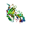
| ||||||||
|---|---|---|---|---|---|---|---|---|---|
| 1 |
| ||||||||
| Unit cell |
|
-Components
| #1: Protein | Mass: 31860.824 Da / Num. of mol.: 1 / Mutation: H269L Source method: isolated from a genetically manipulated source Source: (gene. exp.) Ebola virus sp. / Genus: Ebola-like viruses / Strain: ZAIRE / Description: EBOLA VIRUS RNA GENOME / Plasmid: PRSET / Production host:  Escherichia coli (E. coli) / References: UniProt: Q913A4, UniProt: Q77DJ6*PLUS Escherichia coli (E. coli) / References: UniProt: Q913A4, UniProt: Q77DJ6*PLUS |
|---|---|
| #2: Water | ChemComp-HOH / Water Mass: 18.015 Da / Num. of mol.: 120 / Source method: isolated from a natural source / Formula: H2O Mass: 18.015 Da / Num. of mol.: 120 / Source method: isolated from a natural source / Formula: H2O |
-Experimental details
-Experiment
| Experiment | Method: X-RAY DIFFRACTION / Number of used crystals: 1 |
|---|
- Sample preparation
| Crystal | Density Matthews: 2.25 Å3/Da / Density % sol: 45.41 % | ||||||||||||||||||||||||||||||
|---|---|---|---|---|---|---|---|---|---|---|---|---|---|---|---|---|---|---|---|---|---|---|---|---|---|---|---|---|---|---|---|
| Crystal grow | Temperature: 298 K / Method: vapor diffusion, hanging drop / pH: 8.5 Details: Tris, PEG 750 monomethylether, magnesium chloride, beta-octylglucoside , pH 8.5, VAPOR DIFFUSION, HANGING DROP, temperature 298K | ||||||||||||||||||||||||||||||
| Crystal grow | *PLUS Details: Dessen, A., (2000) Acta Crystallogr., Sect.D, 56, 758. | ||||||||||||||||||||||||||||||
| Components of the solutions | *PLUS
|
-Data collection
| Diffraction | Mean temperature: 100 K |
|---|---|
| Diffraction source | Source: SYNCHROTRON / Site: ESRF  / Beamline: BM14 / Wavelength: 0.8855 / Beamline: BM14 / Wavelength: 0.8855 |
| Detector | Type: MARRESEARCH / Detector: CCD / Date: Nov 8, 1999 |
| Radiation | Protocol: SINGLE WAVELENGTH / Monochromatic (M) / Laue (L): M / Scattering type: x-ray |
| Radiation wavelength | Wavelength: 0.8855 Å / Relative weight: 1 |
| Reflection | Resolution: 2→25 Å / Num. all: 19062 / Num. obs: 17806 / % possible obs: 93.7 % / Observed criterion σ(F): 0 / Observed criterion σ(I): 0 / Redundancy: 8.7 % / Biso Wilson estimate: 24.59 Å2 / Rmerge(I) obs: 0.039 / Net I/σ(I): 10.6 |
| Reflection shell | Resolution: 2→2.05 Å / Redundancy: 3.8 % / Rmerge(I) obs: 0.224 / Num. unique all: 1170 / % possible all: 86.6 |
| Reflection | *PLUS Num. measured all: 166358 |
| Reflection shell | *PLUS % possible obs: 86.8 % / Mean I/σ(I) obs: 3.8 |
- Processing
| Software |
| |||||||||||||||||||||||||
|---|---|---|---|---|---|---|---|---|---|---|---|---|---|---|---|---|---|---|---|---|---|---|---|---|---|---|
| Refinement | Method to determine structure: MAD / Resolution: 2→25 Å / σ(F): 0 / σ(I): 0 / Stereochemistry target values: Engh & Huber
| |||||||||||||||||||||||||
| Refinement step | Cycle: LAST / Resolution: 2→25 Å
| |||||||||||||||||||||||||
| Refine LS restraints |
| |||||||||||||||||||||||||
| Software | *PLUS Name: CNS / Version: 0.9 / Classification: refinement | |||||||||||||||||||||||||
| Refinement | *PLUS Highest resolution: 2 Å / Lowest resolution: 25 Å / σ(F): 0 / % reflection Rfree: 10 % / Rfactor obs: 0.22 / Rfactor Rfree: 0.25 / Rfactor Rwork: 0.22 | |||||||||||||||||||||||||
| Solvent computation | *PLUS | |||||||||||||||||||||||||
| Displacement parameters | *PLUS Biso mean: 28.3 Å2 | |||||||||||||||||||||||||
| LS refinement shell | *PLUS Rfactor Rfree: 0.337 / Rfactor obs: 0.278 |
