 Movie
Movie Controller

[English] 日本語
 Yorodumi
- PDB-1eqz: X-RAY STRUCTURE OF THE NUCLEOSOME CORE PARTICLE AT 2.5 A RESOLUTION -
+ Open data

- Basic information
| Entry | Database: PDB / ID: 1eqz | ||||||
|---|---|---|---|---|---|---|---|
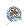
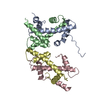
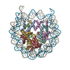
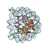
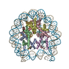
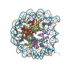
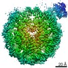
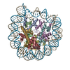
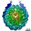
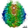
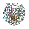
| Title | X-RAY STRUCTURE OF THE NUCLEOSOME CORE PARTICLE AT 2.5 A RESOLUTION | ||||||
 Components Components |
| ||||||
 Keywords Keywords |  STRUCTURAL PROTEIN/DNA / NUCLEOSOME / NUCLEOSOME CORE PARTICLE / HISTONE / MICROGRAVITY HISTONE OCTAMER / DNA PALINDROME / DNA PROTEIN COMPLEX / CHROMATIN / CHROMOSOMAL PROTEIN / HISTONE FOLD / BENT DNA / STRUCTURAL PROTEIN-DNA COMPLEX STRUCTURAL PROTEIN/DNA / NUCLEOSOME / NUCLEOSOME CORE PARTICLE / HISTONE / MICROGRAVITY HISTONE OCTAMER / DNA PALINDROME / DNA PROTEIN COMPLEX / CHROMATIN / CHROMOSOMAL PROTEIN / HISTONE FOLD / BENT DNA / STRUCTURAL PROTEIN-DNA COMPLEX | ||||||
| Function / homology |  Function and homology information Function and homology informationPKMTs methylate histone lysines / HDMs demethylate histones / RMTs methylate histone arginines / SUMOylation of chromatin organization proteins / Condensation of Prophase Chromosomes / Nonhomologous End-Joining (NHEJ) / G2/M DNA damage checkpoint / Metalloprotease DUBs / Processing of DNA double-strand break ends / Recruitment and ATM-mediated phosphorylation of repair and signaling proteins at DNA double strand breaks ...PKMTs methylate histone lysines / HDMs demethylate histones / RMTs methylate histone arginines / SUMOylation of chromatin organization proteins / Condensation of Prophase Chromosomes / Nonhomologous End-Joining (NHEJ) / G2/M DNA damage checkpoint / Metalloprotease DUBs / Processing of DNA double-strand break ends / Recruitment and ATM-mediated phosphorylation of repair and signaling proteins at DNA double strand breaks / Interleukin-7 signaling / Chromatin modifying enzymes / HATs acetylate histones / Ub-specific processing proteases / PRC2 methylates histones and DNA / Oxidative Stress Induced Senescence / B-WICH complex positively regulates rRNA expression / Transcriptional regulation by small RNAs / Assembly of the ORC complex at the origin of replication / RNA Polymerase I Promoter Opening / RNA Polymerase I Promoter Escape / RUNX1 regulates genes involved in megakaryocyte differentiation and platelet function / Estrogen-dependent gene expression / Deposition of new CENPA-containing nucleosomes at the centromere / Factors involved in megakaryocyte development and platelet production / nucleosome assembly / structural constituent of chromatin / nucleosome / protein heterodimerization activity / protein-containing complex binding / DNA binding / nucleoplasm / nucleusSimilarity search - Function | ||||||
| Biological species |  Gallus gallus (chicken) Gallus gallus (chicken) | ||||||
| Method | X-RAY DIFFRACTION / SYNCHROTRON / SIR, molecular replacement / Resolution: 2.5 Å | ||||||
 Authors Authors | Hanson, B.L. / Harp, J.M. / Timm, D.E. / Bunick, G.J. | ||||||
 Citation Citation | Journal: Acta Crystallogr.,Sect.D / Year: 2000 Title: Asymmetries in the nucleosome core particle at 2.5 A resolution. Authors: Harp, J.M. / Hanson, B.L. / Timm, D.E. / Bunick, G.J. #1: Journal: Acta Crystallogr.,Sect.D / Year: 1996Title: X-Ray Diffraction Analysis of Crystals Containing Two Fold Symmetric Nucleosome Core Particles Authors: Harp, J.M. / Uberbacher, E.C. / Roberson, A. / Palmer, E. / Gewiess, A. / Bunick, G.J. #2: Journal: Thesis / Year: 1985Title: Structural Analysis of Members of a Repeated DNA Family in Primates Authors: Hauser, L.J. | ||||||
| History |
|
- Structure visualization
| Structure viewer | Molecule: MolmilJmol/JSmol |
|---|
- Downloads & links
-Download
| PDBx/mmCIF format | 1eqz.cif.gz | 354.4 KB | Display | PDBx/mmCIF format |
|---|---|---|---|---|
| PDB format | pdb1eqz.ent.gz | 272.4 KB | Display | PDB format |
| PDBx/mmJSON format | 1eqz.json.gz | Tree view | PDBx/mmJSON format | |
| Others |  Other downloads Other downloads |
-Validation report
| Arichive directory | https://data.pdbj.org/pub/pdb/validation_reports/eq/1eqzftp://data.pdbj.org/pub/pdb/validation_reports/eq/1eqz | HTTPS FTP |
|---|
-Related structure data

-Links
 PDBj
PDBj


- Assembly
| Deposited unit | 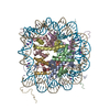
| ||||||||||
|---|---|---|---|---|---|---|---|---|---|---|---|
| 1 |
| ||||||||||
| Unit cell |
|
-Components
-DNA chain , 1 types, 2 molecules IJ
| #1: DNA chain | Mass: 45054.844 Da / Num. of mol.: 2 / Source method: obtained synthetically Details: 146 BP DNA PALINDROME BASED ON 5' HALF OF NUCLEOSOME PHASING SEQUENCE OF ALPHA SATELLITE DNA FROM HUMAN X CHROMOSOME BAM H1 REPEAT |
|---|
-PROTEIN (HISTONE ... , 4 types, 8 molecules AEBFCGDH
| #2: Protein | Mass: 13969.363 Da / Num. of mol.: 2 / Source method: isolated from a natural source / Details: SUBUNIT OF SALT-EXTRACTED HISTONE OCTAMER / Source: (natural) Gallus gallus (chicken) / Organelle: ERYTHROCYTE NUCLEUS / Tissue: BLOOD / References: UniProt: P02263#3: Protein | Mass: 13953.251 Da / Num. of mol.: 2 / Source method: isolated from a natural source / Details: SUBUNIT OF SALT-EXTRACTED HISTONE OCTAMER / Source: (natural) Gallus gallus (chicken) / Organelle: ERYTHROCYTE NUCLEUS / Tissue: BLOOD / References: UniProt: P02279, UniProt: P0C1H5*PLUS#4: Protein | Mass: 15421.101 Da / Num. of mol.: 2 / Source method: isolated from a natural source / Details: SUBUNIT OF SALT-EXTRACTED HISTONE OCTAMER / Source: (natural) Gallus gallus (chicken) / Organelle: ERYTHROCYTE NUCLEUS / Tissue: BLOOD / References: UniProt: P84229#5: Protein | Mass: 11394.426 Da / Num. of mol.: 2 / Source method: isolated from a natural source / Details: SUBUNIT OF SALT-EXTRACTED HISTONE OCTAMER / Source: (natural) Gallus gallus (chicken) / Organelle: ERYTHROCYTE NUCLEUS / Tissue: BLOOD / References: UniProt: P62801 |
|---|
-Non-polymers , 5 types, 374 molecules 


| #6: Chemical | ChemComp-K /  Mass: 39.098 Da / Num. of mol.: 7 / Source method: obtained synthetically / Formula: K Mass: 39.098 Da / Num. of mol.: 7 / Source method: obtained synthetically / Formula: K#7: Chemical | ChemComp-MN /  Mass: 54.938 Da / Num. of mol.: 15 / Source method: obtained synthetically / Formula: Mn Mass: 54.938 Da / Num. of mol.: 15 / Source method: obtained synthetically / Formula: Mn#8: Chemical | Chloride Mass: 35.453 Da / Num. of mol.: 2 / Source method: obtained synthetically / Formula: Cl Mass: 35.453 Da / Num. of mol.: 2 / Source method: obtained synthetically / Formula: Cl#9: Chemical | ChemComp-CAC / | Cacodylic acid Mass: 136.989 Da / Num. of mol.: 1 / Source method: obtained synthetically / Formula: C2H6AsO2 Mass: 136.989 Da / Num. of mol.: 1 / Source method: obtained synthetically / Formula: C2H6AsO2#10: Water | ChemComp-HOH / | WaterMass: 18.015 Da / Num. of mol.: 349 / Source method: isolated from a natural source / Formula: H2O |
|---|
-Experimental details
-Experiment
| Experiment | Method: X-RAY DIFFRACTION / Number of used crystals: 1 |
|---|
- Sample preparation
| Crystal | Density Matthews: 2.62 Å3/Da / Density % sol: 53.06 % | |||||||||||||||||||||||||||||||||||||||||||||||||||||||||||||||
|---|---|---|---|---|---|---|---|---|---|---|---|---|---|---|---|---|---|---|---|---|---|---|---|---|---|---|---|---|---|---|---|---|---|---|---|---|---|---|---|---|---|---|---|---|---|---|---|---|---|---|---|---|---|---|---|---|---|---|---|---|---|---|---|---|
| Crystal grow | Method: vapor diffusion, hanging drop / pH: 6 / Details: pH 6.0, VAPOR DIFFUSION, HANGING DROP | |||||||||||||||||||||||||||||||||||||||||||||||||||||||||||||||
| Crystal grow | *PLUS Method: unknown | |||||||||||||||||||||||||||||||||||||||||||||||||||||||||||||||
| Components of the solutions | *PLUS
|
-Data collection
| Diffraction | Mean temperature: 100 K |
|---|---|
| Diffraction source | Source: SYNCHROTRON / Site: NSLS  / Beamline: X12C / Wavelength: 1.003 / Beamline: X12C / Wavelength: 1.003 |
| Detector | Type: MARRESEARCH / Detector: IMAGE PLATE / Date: Apr 18, 1996 |
| Radiation | Protocol: SINGLE WAVELENGTH / Monochromatic (M) / Laue (L): M / Scattering type: x-ray |
| Radiation wavelength | Wavelength: 1.003 Å / Relative weight: 1 |
| Reflection | Resolution: 2.5→25.4 Å / Num. all: 73319 / Num. obs: 73319 / % possible obs: 98.7 % / Observed criterion σ(I): 0 / Redundancy: 8.3 % / Biso Wilson estimate: 42.7 Å2 / Rsym value: 0.053 |
| Reflection | *PLUS Lowest resolution: 25 Å / Num. obs: 72200 / Num. measured all: 605950 / Rmerge(I) obs: 0.045 |
- Processing
| Software |
| ||||||||||||||||||||||||||||||||||||||||||||||||||||||||||||||||||||||||||||||||
|---|---|---|---|---|---|---|---|---|---|---|---|---|---|---|---|---|---|---|---|---|---|---|---|---|---|---|---|---|---|---|---|---|---|---|---|---|---|---|---|---|---|---|---|---|---|---|---|---|---|---|---|---|---|---|---|---|---|---|---|---|---|---|---|---|---|---|---|---|---|---|---|---|---|---|---|---|---|---|---|---|---|
| Refinement | Method to determine structure: SIR, molecular replacement Starting model: 2HIO, 1AOI Resolution: 2.5→25.34 Å / Rfactor Rfree error: 0.003 / Data cutoff high absF: 2059998.46 / Data cutoff low absF: 0 / Isotropic thermal model: RESTRAINED / Cross valid method: THROUGHOUT / σ(F): 0
| ||||||||||||||||||||||||||||||||||||||||||||||||||||||||||||||||||||||||||||||||
| Solvent computation | Solvent model: FLAT MODEL / Bsol: 68.68 Å2 / ksol: 0.393 e/Å3 | ||||||||||||||||||||||||||||||||||||||||||||||||||||||||||||||||||||||||||||||||
| Displacement parameters | Biso mean: 53.2 Å2
| ||||||||||||||||||||||||||||||||||||||||||||||||||||||||||||||||||||||||||||||||
| Refine analyze |
| ||||||||||||||||||||||||||||||||||||||||||||||||||||||||||||||||||||||||||||||||
| Refinement step | Cycle: LAST / Resolution: 2.5→25.34 Å
| ||||||||||||||||||||||||||||||||||||||||||||||||||||||||||||||||||||||||||||||||
| Refine LS restraints |
| ||||||||||||||||||||||||||||||||||||||||||||||||||||||||||||||||||||||||||||||||
| Xplor file |
| ||||||||||||||||||||||||||||||||||||||||||||||||||||||||||||||||||||||||||||||||
| Software | *PLUS Name: CNS / Version: 1 / Classification: refinement | ||||||||||||||||||||||||||||||||||||||||||||||||||||||||||||||||||||||||||||||||
| Refine LS restraints | *PLUS
|
