 Movie
Movie Controller

[English] 日本語
 Yorodumi
- PDB-1eo8: INFLUENZA VIRUS HEMAGGLUTININ COMPLEXED WITH A NEUTRALIZING ANTIBODY -
+ Open data

- Basic information
| Entry | Database: PDB / ID: 1eo8 | |||||||||
|---|---|---|---|---|---|---|---|---|---|---|
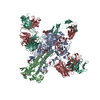
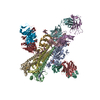
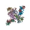
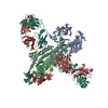
| Title | INFLUENZA VIRUS HEMAGGLUTININ COMPLEXED WITH A NEUTRALIZING ANTIBODY | |||||||||
 Components Components |
| |||||||||
 Keywords Keywords |  Viral protein/Immune system / COMPLEX (HEMAGGLUTININ-IMMMUNOGLOBULIN) / HEMAGGLUTININ / IMMUNOGLOBULIN / VIRAL PROTEIN / IMMUNE SYSTEM COMPLEX / Viral protein-Immune system COMPLEX Viral protein/Immune system / COMPLEX (HEMAGGLUTININ-IMMMUNOGLOBULIN) / HEMAGGLUTININ / IMMUNOGLOBULIN / VIRAL PROTEIN / IMMUNE SYSTEM COMPLEX / Viral protein-Immune system COMPLEX | |||||||||
| Function / homology |  Function and homology informationviral budding from plasma membrane / clathrin-dependent endocytosis of virus by host cell / host cell surface receptor binding / fusion of virus membrane with host plasma membrane / fusion of virus membrane with host endosome membrane / viral envelope / virion attachment to host cell / host cell plasma membrane / virion membrane / membrane Function and homology informationviral budding from plasma membrane / clathrin-dependent endocytosis of virus by host cell / host cell surface receptor binding / fusion of virus membrane with host plasma membrane / fusion of virus membrane with host endosome membrane / viral envelope / virion attachment to host cell / host cell plasma membrane / virion membrane / membraneSimilarity search - Function | |||||||||
| Biological species |   Influenza A virus Influenza A virus Mus musculus (house mouse) Mus musculus (house mouse) | |||||||||
| Method | X-RAY DIFFRACTION / SYNCHROTRON / MOLECULAR REPLACEMENT / Resolution: 2.8 Å | |||||||||
 Authors Authors | Fleury, D. / Gigant, B. / Daniels, R.S. / Skehel, J.J. / Knossow, M. / Bizebard, T. | |||||||||
 Citation Citation | Journal: Proteins / Year: 2000 Title: Structural evidence for recognition of a single epitope by two distinct antibodies. Authors: Fleury, D. / Daniels, R.S. / Skehel, J.J. / Knossow, M. / Bizebard, T. #1: Journal: Proteins / Year: 1995Title: Crystallization and Preliminary X-Ray Diffraction Studies of Complexes between an Influenza Hemagglutinin and Fab Fragments of Two Different Monoclonal Antibodies Authors: Gigant, B. / Fleury, D. / Bizebard, T. / Skehel, J.J. / Knossow, M. #2: Journal: Nature / Year: 1981Title: Structure of the Haemagglutinin Membrane Glycoprotein of Influenza Virus at 3 A Resolution Authors: Wilson, I.A. / Skehel, J.J. / Wiley, D.C. | |||||||||
| History |
|
- Structure visualization
| Structure viewer | Molecule: MolmilJmol/JSmol |
|---|
- Downloads & links
-Download
| PDBx/mmCIF format | 1eo8.cif.gz | 192.5 KB | Display | PDBx/mmCIF format |
|---|---|---|---|---|
| PDB format | pdb1eo8.ent.gz | 155.2 KB | Display | PDB format |
| PDBx/mmJSON format | 1eo8.json.gz | Tree view | PDBx/mmJSON format | |
| Others |  Other downloads Other downloads |
-Validation report
| Arichive directory | https://data.pdbj.org/pub/pdb/validation_reports/eo/1eo8ftp://data.pdbj.org/pub/pdb/validation_reports/eo/1eo8 | HTTPS FTP |
|---|
-Related structure data
| Similar structure data |
|---|


-Links
 PDBj
PDBj


- Assembly
| Deposited unit | 
| ||||||||
|---|---|---|---|---|---|---|---|---|---|
| 1 | 
| ||||||||
| Unit cell |
| ||||||||
| Details | THERE IS ONE MONOMER OF THE TRIMERIC HEMAGGLUTININ MOLECULE IN THE ASYMMETRIC UNIT, AND EACH MONOMER IS COMPLEXED WITH ONE FAB FRAGMENT. THE MONOMER OF HEMAGGLUTININ CONSISTS OF TWO CHAINS, IDENTIFIED AS HA1 AND HA2. CHAINS HA1 AND HA2 HAVE BEEN ASSIGNED CHAIN IDENTIFIERS *A* AND *B*, RESPECTIVELY. IN THE VIRUS, CHAIN HA1 CONSISTS OF 328 RESIDUES AND CHAIN HA2 CONSISTS OF 220 RESIDUES. HEMAGGLUTININ MAY BE SOLUBILIZED FROM THE VIRAL MEMBRANE BY BROMELAIN DIGESTION, WHICH REMOVES THE C-TERMINAL HYDROPHOBIC (ANCHORING) DOMAIN FROM CHAIN HA2. AFTER BROMELAIN DIGESTION CHAIN HA2 CONSISTS OF 175 RESIDUES, AS PRESENTED IN THIS ENTRY. |
-Components
-HEMAGGLUTININ ... , 2 types, 2 molecules AB
| #1: Protein | Mass: 36065.457 Da / Num. of mol.: 1 / Fragment: BROMELAIN RELEASED FRAGMENT / Source method: isolated from a natural source Details: A RECOMBINANT INFLUENZA STRAIN CONTAINING A/AICHI/68 (H3N2) HEMAGGLUTININ Source: (natural) Influenza A virus (A/X-31(H3N2)) / Genus: Influenzavirus A / Species: Influenza A virus / Strain: X31 / References: UniProt: P03437 |
|---|---|
| #2: Protein | Mass: 20212.350 Da / Num. of mol.: 1 / Fragment: BROMELAIN RELEASED FRAGMENT / Source method: isolated from a natural source Details: A RECOMBINANT INFLUENZA STRAIN CONTAINING A/AICHI/68 (H3N2) HEMAGGLUTININ Source: (natural) Influenza A virus (A/X-31(H3N2)) / Genus: Influenzavirus A / Species: Influenza A virus / Strain: X31 / References: UniProt: P03437 |
-Antibody , 2 types, 2 molecules LH
| #3: Antibody | Mass: 23097.604 Da / Num. of mol.: 1 / Fragment: FAB FRAGMENT OF ANTIBODY BH151 / Source method: isolated from a natural source / Source: (natural) Mus musculus (house mouse) / Cell: HYBRIDROMAS / Strain: BALB/C / References: GenBank: 7159941 |
|---|---|
| #4: Antibody | Mass: 23407.254 Da / Num. of mol.: 1 / Fragment: FAB FRAGMENT OF ANTIBODY BH151 / Source method: isolated from a natural source / Source: (natural) Mus musculus (house mouse) / Cell: HYBRIDROMAS / Strain: BALB/C / References: GenBank: 7159939 |
-Sugars , 3 types, 4 molecules 
| #5: Polysaccharide | alpha-D-mannopyranose-(1-4)-2-acetamido-2-deoxy-beta-D-glucopyranose-(1-4)-2-acetamido-2-deoxy-beta- ...alpha-D-mannopyranose-(1-4)-2-acetamido-2-deoxy-beta-D-glucopyranose-(1-4)-2-acetamido-2-deoxy-beta-D-glucopyranose / Mass: 586.542 Da / Num. of mol.: 1 Source method: isolated from a genetically manipulated source |
|---|---|
| #6: Polysaccharide | 2-acetamido-2-deoxy-beta-D-glucopyranose-(1-4)-2-acetamido-2-deoxy-beta-D-glucopyranose / Mass: 424.401 Da / Num. of mol.: 1 Source method: isolated from a genetically manipulated source |
| #7: Sugar | N-Acetylglucosamine Type: D-saccharide, beta linking / Mass: 221.208 Da / Num. of mol.: 2 Type: D-saccharide, beta linking / Mass: 221.208 Da / Num. of mol.: 2Source method: isolated from a genetically manipulated source Formula: C8H15NO6 |
-Non-polymers , 1 types, 109 molecules 
| #8: Water | ChemComp-HOH / WaterMass: 18.015 Da / Num. of mol.: 109 / Source method: isolated from a natural source / Formula: H2O |
|---|
-Experimental details
-Experiment
| Experiment | Method: X-RAY DIFFRACTION / Number of used crystals: 1 |
|---|
- Sample preparation
| Crystal | Density Matthews: 3.47 Å3/Da / Density % sol: 64.54 % | ||||||||||||||||||||||||||||||||||||||||||
|---|---|---|---|---|---|---|---|---|---|---|---|---|---|---|---|---|---|---|---|---|---|---|---|---|---|---|---|---|---|---|---|---|---|---|---|---|---|---|---|---|---|---|---|
| Crystal grow | Temperature: 293 K / Method: vapor diffusion, hanging drop / pH: 6 Details: 28%(w:v) PEG 600, 100 mM Sodium Phosphate, 150 mM NaCl, 0.05% NaN3 , pH 6.0, VAPOR DIFFUSION, HANGING DROP, temperature 293K | ||||||||||||||||||||||||||||||||||||||||||
| Crystal grow | *PLUS Temperature: 18 ℃ / pH: 8.5 Details: Gigant, B., (1995) Proteins: Struct., Funct., Genet., 23, 115. | ||||||||||||||||||||||||||||||||||||||||||
| Components of the solutions | *PLUS
|
-Data collection
| Diffraction | Mean temperature: 100 K |
|---|---|
| Diffraction source | Source: SYNCHROTRON / Site: ESRF  / Beamline: ID2 / Wavelength: 1 / Beamline: ID2 / Wavelength: 1 |
| Detector | Type: MARRESEARCH / Detector: IMAGE PLATE / Date: Nov 10, 1995 |
| Radiation | Protocol: SINGLE WAVELENGTH / Monochromatic (M) / Laue (L): M / Scattering type: x-ray |
| Radiation wavelength | Wavelength: 1 Å / Relative weight: 1 |
| Reflection | Resolution: 2.8→25 Å / Num. all: 186998 / Num. obs: 49210 / % possible obs: 99 % / Observed criterion σ(F): 0 / Observed criterion σ(I): 0 / Redundancy: 3.8 % / Rmerge(I) obs: 0.068 / Rsym value: 0.068 / Net I/σ(I): 7.5 |
| Reflection shell | Resolution: 2.8→2.85 Å / Redundancy: 3 % / Rmerge(I) obs: 0.336 / Rsym value: 0.336 / % possible all: 99 |
| Reflection | *PLUS Num. measured all: 186998 |
| Reflection shell | *PLUS % possible obs: 99 % |
- Processing
| Software |
| |||||||||||||||
|---|---|---|---|---|---|---|---|---|---|---|---|---|---|---|---|---|
| Refinement | Method to determine structure: MOLECULAR REPLACEMENT / Resolution: 2.8→7 Å / Cross valid method: RFREE / σ(F): 2
| |||||||||||||||
| Refinement step | Cycle: LAST / Resolution: 2.8→7 Å
| |||||||||||||||
| Refine LS restraints |
| |||||||||||||||
| Software | *PLUS Name: X-PLOR / Version: 3.843 / Classification: refinement | |||||||||||||||
| Refinement | *PLUS Highest resolution: 2.8 Å / Lowest resolution: 7 Å / σ(F): 2 / % reflection Rfree: 5 % / Rfactor obs: 0.196 | |||||||||||||||
| Solvent computation | *PLUS | |||||||||||||||
| Displacement parameters | *PLUS | |||||||||||||||
| Refine LS restraints | *PLUS Type: x_angle_deg / Dev ideal: 1.9 |
