 Movie
Movie Controller


+ Open data

- Basic information
| Entry | Database: PDB / ID: 1ekr | ||||||
|---|---|---|---|---|---|---|---|
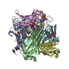
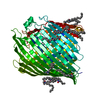
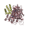
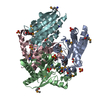
| Title | MOAC PROTEIN FROM E. COLI | ||||||
 Components Components | MOLYBDENUM COFACTOR BIOSYNTHESIS PROTEIN C | ||||||
 Keywords Keywords |  TRANSLATION / MoaC / Molybdenum cofactor (Moco) / Moco biosynthesis / Moco deficiency TRANSLATION / MoaC / Molybdenum cofactor (Moco) / Moco biosynthesis / Moco deficiency | ||||||
| Function / homology |  Function and homology informationcyclic pyranopterin monophosphate synthase / cyclic pyranopterin monophosphate synthase activity / Mo-molybdopterin cofactor biosynthetic process / protein-containing complex / identical protein binding Function and homology informationcyclic pyranopterin monophosphate synthase / cyclic pyranopterin monophosphate synthase activity / Mo-molybdopterin cofactor biosynthetic process / protein-containing complex / identical protein bindingSimilarity search - Function | ||||||
| Biological species |  Escherichia coli (E. coli) Escherichia coli (E. coli) | ||||||
| Method | X-RAY DIFFRACTION / SYNCHROTRON / Resolution: 2 Å | ||||||
 Authors Authors | Schindelin, H. / Liu, M.T.W. / Wuebbens, M.M. / Rajagopalan, K.V. | ||||||
 Citation Citation | Journal: Structure Fold.Des. / Year: 2000 Title: Insights into molybdenum cofactor deficiency provided by the crystal structure of the molybdenum cofactor biosynthesis protein MoaC. Authors: Wuebbens, M.M. / Liu, M.T. / Rajagopalan, K. / Schindelin, H. | ||||||
| History |
|
- Structure visualization
| Structure viewer | Molecule: MolmilJmol/JSmol |
|---|
- Downloads & links
-Download
| PDBx/mmCIF format | 1ekr.cif.gz | 40.8 KB | Display | PDBx/mmCIF format |
|---|---|---|---|---|
| PDB format | pdb1ekr.ent.gz | 28.9 KB | Display | PDB format |
| PDBx/mmJSON format | 1ekr.json.gz | Tree view | PDBx/mmJSON format | |
| Others |  Other downloads Other downloads |
-Validation report
| Arichive directory | https://data.pdbj.org/pub/pdb/validation_reports/ek/1ekrftp://data.pdbj.org/pub/pdb/validation_reports/ek/1ekr | HTTPS FTP |
|---|
-Related structure data


-Links
 PDBj
PDBj- Assembly
| Deposited unit | 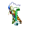
| ||||||||
|---|---|---|---|---|---|---|---|---|---|
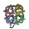
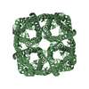
| 1 | x 6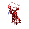
| ||||||||
| Unit cell |
| ||||||||
| Details | The biological assembly is a hexamer with 32 symmetry generated from chain A by crystallographic symmetry operations |
-Components
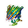
| #1: Protein | Mass: 17477.277 Da / Num. of mol.: 1 / Mutation: S2A Source method: isolated from a genetically manipulated source Source: (gene. exp.) Escherichia coli (E. coli) / Plasmid: PET 15B / Cellular location (production host): CYTOSOL / Production host: Escherichia coli (E. coli) / References: UniProt: P0A738 |
|---|---|
| #2: Water | ChemComp-HOH / Water Mass: 18.015 Da / Num. of mol.: 41 / Source method: isolated from a natural source / Formula: H2O Mass: 18.015 Da / Num. of mol.: 41 / Source method: isolated from a natural source / Formula: H2O |
-Experimental details
-Experiment
| Experiment | Method: X-RAY DIFFRACTION / Number of used crystals: 1 |
|---|
- Sample preparation
| Crystal | Density Matthews: 2.15 Å3/Da / Density % sol: 42.78 % | ||||||||||||||||||||||||||||||
|---|---|---|---|---|---|---|---|---|---|---|---|---|---|---|---|---|---|---|---|---|---|---|---|---|---|---|---|---|---|---|---|
| Crystal grow | Temperature: 295 K / Method: vapor diffusion, sitting drop / pH: 7.5 Details: PEG 400, CALCIUM CHLORIDE, HEPES, pH 7.5, VAPOR DIFFUSION, SITTING DROP, temperature 22K | ||||||||||||||||||||||||||||||
| Crystal | *PLUS Density % sol: 42 % | ||||||||||||||||||||||||||||||
| Crystal grow | *PLUS Method: vapor diffusion | ||||||||||||||||||||||||||||||
| Components of the solutions | *PLUS
|
-Data collection
| Diffraction | Mean temperature: 95 K |
|---|---|
| Diffraction source | Source: SYNCHROTRON / Site: NSLS  / Beamline: X26C / Wavelength: 1.1 / Beamline: X26C / Wavelength: 1.1 |
| Detector | Type: ADSC QUANTUM 4 / Detector: CCD / Date: Mar 15, 1998 |
| Radiation | Protocol: SINGLE WAVELENGTH / Monochromatic (M) / Laue (L): M / Scattering type: x-ray |
| Radiation wavelength | Wavelength: 1.1 Å / Relative weight: 1 |
| Reflection | Resolution: 1.95→20 Å / Num. all: 57577 / Num. obs: 11190 / % possible obs: 95.7 % / Observed criterion σ(F): 0 / Observed criterion σ(I): 0 / Redundancy: 5.1 % / Biso Wilson estimate: 48 Å2 / Rmerge(I) obs: 0.063 / Net I/σ(I): 29.1 |
| Reflection shell | Resolution: 1.95→2.02 Å / Redundancy: 3.9 % / Rmerge(I) obs: 0.497 / Num. unique all: 1083 / % possible all: 95.7 |
| Reflection | *PLUS Highest resolution: 2 Å / Lowest resolution: 50 Å / Redundancy: 5.2 % |
| Reflection shell | *PLUS % possible obs: 97.9 % / Rmerge(I) obs: 0.365 / Mean I/σ(I) obs: 3.5 |
- Processing
| Software |
| |||||||||||||||||||||||||||||||||||||||
|---|---|---|---|---|---|---|---|---|---|---|---|---|---|---|---|---|---|---|---|---|---|---|---|---|---|---|---|---|---|---|---|---|---|---|---|---|---|---|---|---|
| Refinement | Resolution: 2→20 Å / σ(F): 0 / σ(I): 0 / Stereochemistry target values: Lamzin Details: Partial structure factors for bulk solvent were calculated in XPLOR and incorporated into REFMAC
| |||||||||||||||||||||||||||||||||||||||
| Refinement step | Cycle: LAST / Resolution: 2→20 Å
| |||||||||||||||||||||||||||||||||||||||
| Refine LS restraints |
| |||||||||||||||||||||||||||||||||||||||
| Software | *PLUS Name: REFMAC / Classification: refinement | |||||||||||||||||||||||||||||||||||||||
| Refinement | *PLUS Rfactor Rfree: 0.252 / Rfactor Rwork: 0.219 | |||||||||||||||||||||||||||||||||||||||
| Solvent computation | *PLUS | |||||||||||||||||||||||||||||||||||||||
| Displacement parameters | *PLUS | |||||||||||||||||||||||||||||||||||||||
| Refine LS restraints | *PLUS
|
