 Movie
Movie Controller


+ Open data

- Basic information
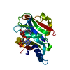
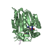
| Entry | Database: PDB / ID: 1ej4 | |||||||||
|---|---|---|---|---|---|---|---|---|---|---|
| Title | COCRYSTAL STRUCTURE OF EIF4E/4E-BP1 PEPTIDE | |||||||||
 Components Components |
| |||||||||
 Keywords Keywords |  TRANSLATION / eIF4E/4E-BP/7-methyl-GDP TRANSLATION / eIF4E/4E-BP/7-methyl-GDP | |||||||||
| Function / homology |  Function and homology information Function and homology informationActivation of the mRNA upon binding of the cap-binding complex and eIFs, and subsequent binding to 43S / Transport of the SLBP independent Mature mRNA / Transport of the SLBP Dependant Mature mRNA / Transport of Mature mRNA Derived from an Intronless Transcript / Deadenylation of mRNA / Translation initiation complex formation / Ribosomal scanning and start codon recognition / ISG15 antiviral mechanism / L13a-mediated translational silencing of Ceruloplasmin expression / GTP hydrolysis and joining of the 60S ribosomal subunit ...Activation of the mRNA upon binding of the cap-binding complex and eIFs, and subsequent binding to 43S / Transport of the SLBP independent Mature mRNA / Transport of the SLBP Dependant Mature mRNA / Transport of Mature mRNA Derived from an Intronless Transcript / Deadenylation of mRNA / Translation initiation complex formation / Ribosomal scanning and start codon recognition / ISG15 antiviral mechanism / L13a-mediated translational silencing of Ceruloplasmin expression / GTP hydrolysis and joining of the 60S ribosomal subunit / mTORC1-mediated signalling / Activation of the mRNA upon binding of the cap-binding complex and eIFs, and subsequent binding to 43S / eukaryotic initiation factor 4G binding / eukaryotic initiation factor 4E binding / regulation of translation at postsynapse, modulating synaptic transmission / chromatoid body / eukaryotic translation initiation factor 4F complex / mRNA cap binding / : / RNA 7-methylguanosine cap binding / RISC complex / nuclear export / postsynaptic cytosol / stem cell population maintenance / TOR signaling / mTORC1-mediated signalling / negative regulation of neuron differentiation / behavioral fear response / mRNA export from nucleus / translation initiation factor binding / translation repressor activity / negative regulation of translational initiation / translational initiation / translation initiation factor activity / cellular response to dexamethasone stimulus / positive regulation of mitotic cell cycle / P-body / G1/S transition of mitotic cell cycle / neuron differentiation / cytoplasmic ribonucleoprotein granule / cytoplasmic stress granule / regulation of translation / postsynapse / DNA-binding transcription factor binding / negative regulation of translation / nuclear body / nuclear speck / translation / glutamatergic synapse / perinuclear region of cytoplasm / enzyme binding / protein-containing complex / nucleus / cytosol / cytoplasmSimilarity search - Function | |||||||||
| Biological species |  Mus musculus (house mouse) Mus musculus (house mouse) | |||||||||
| Method | X-RAY DIFFRACTION / Resolution: 2.25 Å | |||||||||
 Authors Authors | Marcotrigiano, J. / Gingras, A.-C. / Sonenberg, N. / Burley, S.K. | |||||||||
 Citation Citation | Journal: Mol.Cell / Year: 1999 Title: Cap-dependent translation initiation in eukaryotes is regulated by a molecular mimic of eIF4G. Authors: Marcotrigiano, J. / Gingras, A.C. / Sonenberg, N. / Burley, S.K. | |||||||||
| History |
|
- Structure visualization
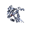
| Structure viewer | Molecule: MolmilJmol/JSmol |
|---|
- Downloads & links
-Download
| PDBx/mmCIF format | 1ej4.cif.gz | 55.5 KB | Display | PDBx/mmCIF format |
|---|---|---|---|---|
| PDB format | pdb1ej4.ent.gz | 39.2 KB | Display | PDB format |
| PDBx/mmJSON format | 1ej4.json.gz | Tree view | PDBx/mmJSON format | |
| Others |  Other downloads Other downloads |
-Validation report
| Arichive directory | https://data.pdbj.org/pub/pdb/validation_reports/ej/1ej4ftp://data.pdbj.org/pub/pdb/validation_reports/ej/1ej4 | HTTPS FTP |
|---|
-Related structure data


-Links
 PDBj
PDBj
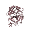
- Assembly
| Deposited unit | 
| ||||||||
|---|---|---|---|---|---|---|---|---|---|
| 1 |
| ||||||||
| Unit cell |
|
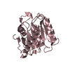
-Components
| #1: Protein | Mass: 22145.113 Da / Num. of mol.: 1 / Fragment: RESIDUES 28-217 Source method: isolated from a genetically manipulated source Source: (gene. exp.) Mus musculus (house mouse) / Plasmid: PET3B / Production host:  Escherichia coli (E. coli) / References: UniProt: P63073 Escherichia coli (E. coli) / References: UniProt: P63073 |
|---|---|
| #2: Protein/peptide | Mass: 1861.240 Da / Num. of mol.: 1 / Fragment: RESIDUES 51-67 / Source method: obtained synthetically Details: THIS PEPTIDE WAS CHEMICALLY SYNTHESIZED. THE SEQUENCE OF THIS PEPTIDE NATURALLY OCCURS IN HUMANS (HOMO SAPIENS) References: GenBank: 4758258, UniProt: Q13541*PLUS |
| #3: Chemical | ChemComp-M7G / 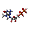  Mass: 458.235 Da / Num. of mol.: 1 / Source method: obtained synthetically / Formula: C11H18N5O11P2 Mass: 458.235 Da / Num. of mol.: 1 / Source method: obtained synthetically / Formula: C11H18N5O11P2 |
| #4: Water | ChemComp-HOH / Water Mass: 18.015 Da / Num. of mol.: 85 / Source method: isolated from a natural source / Formula: H2O Mass: 18.015 Da / Num. of mol.: 85 / Source method: isolated from a natural source / Formula: H2O |
-Experimental details
-Experiment
| Experiment | Method: X-RAY DIFFRACTION / Number of used crystals: 1 |
|---|
- Sample preparation
| Crystal | Density Matthews: 2.34 Å3/Da / Density % sol: 47.5 % | ||||||||||||||||||||
|---|---|---|---|---|---|---|---|---|---|---|---|---|---|---|---|---|---|---|---|---|---|
| Crystal grow | Temperature: 293 K / Method: vapor diffusion, hanging drop / pH: 7.5 Details: PEG4000, pH 7.5, VAPOR DIFFUSION, HANGING DROP, temperature 20K | ||||||||||||||||||||
| Crystal grow | *PLUS | ||||||||||||||||||||
| Components of the solutions | *PLUS
|
-Data collection
| Diffraction | Mean temperature: 100 K |
|---|---|
| Diffraction source | Source: ROTATING ANODE / Type: RIGAKU RU200 / Wavelength: 1.5418 |
| Detector | Type: RIGAKU RAXIS IIC / Detector: IMAGE PLATE / Date: Sep 15, 1997 |
| Radiation | Protocol: SINGLE WAVELENGTH / Monochromatic (M) / Laue (L): M / Scattering type: x-ray |
| Radiation wavelength | Wavelength: 1.5418 Å / Relative weight: 1 |
| Reflection | Resolution: 2.25→30 Å / Num. all: 10817 / % possible obs: 97.4 % / Observed criterion σ(I): 5 / Redundancy: 4 % / Rmerge(I) obs: 0.052 / Net I/σ(I): 24.9 |
| Reflection shell | Resolution: 2.25→2.33 Å / Redundancy: 4 % / Rmerge(I) obs: 0.103 / % possible all: 84.8 |
| Reflection shell | *PLUS % possible obs: 84.8 % |
- Processing
| Software |
| ||||||||||||
|---|---|---|---|---|---|---|---|---|---|---|---|---|---|
| Refinement | Resolution: 2.25→25 Å / σ(F): 2
| ||||||||||||
| Refinement step | Cycle: LAST / Resolution: 2.25→25 Å
| ||||||||||||
| Software | *PLUS Name: 'CNS' / Classification: refinement | ||||||||||||
| Refine LS restraints | *PLUS
|
