 Movie
Movie Controller

[English] 日本語
 Yorodumi
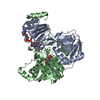
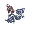
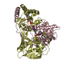
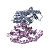
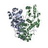
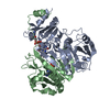
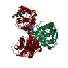
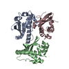
- PDB-1efp: ELECTRON TRANSFER FLAVOPROTEIN (ETF) FROM PARACOCCUS DENITRIFICANS -
+ Open data

- Basic information
| Entry | Database: PDB / ID: 1efp | ||||||
|---|---|---|---|---|---|---|---|
| Title | ELECTRON TRANSFER FLAVOPROTEIN (ETF) FROM PARACOCCUS DENITRIFICANS | ||||||
 Components Components | (PROTEIN (ELECTRON TRANSFER FLAVOPROTEIN)) x 2 | ||||||
 Keywords Keywords |  ELECTRON TRANSPORT / FLAVOPROTEIN / GLUTARIC ACIDEMIA TYPE II ELECTRON TRANSPORT / FLAVOPROTEIN / GLUTARIC ACIDEMIA TYPE II | ||||||
| Function / homology |  Function and homology information Function and homology information | ||||||
| Biological species |  Paracoccus denitrificans (bacteria) Paracoccus denitrificans (bacteria) | ||||||
| Method | X-RAY DIFFRACTION / MOLECULAR REPLACEMENT / Resolution: 2.6 Å | ||||||
 Authors Authors | Roberts, D.L. / Salazar, D. / Fulmer, J.P. / Frerman, F.E. / Kim, J.J.-P. | ||||||
 Citation Citation | Journal: Biochemistry / Year: 1999 Title: Crystal structure of Paracoccus denitrificans electron transfer flavoprotein: structural and electrostatic analysis of a conserved flavin binding domain. Authors: Roberts, D.L. / Salazar, D. / Fulmer, J.P. / Frerman, F.E. / Kim, J.J. #1: Journal: Proc.Natl.Acad.Sci.USA / Year: 1996Title: Three-Dimensional Structure of Human Electron Transfer Flavoprotein to 2.1-A Resolution Authors: Roberts, D.L. / Frerman, F.E. / Kim, J.J. #2: Journal: Protein Sci. / Year: 1995Title: Crystallization and Preliminary X-Ray Analysis of Electron Transfer Flavoproteins from Human and Paracoccus Denitrificans Authors: Roberts, D.L. / Herrick, K.R. / Frerman, F.E. / Kim, J.J. | ||||||
| History |
|
- Structure visualization
| Structure viewer | Molecule: MolmilJmol/JSmol |
|---|
- Downloads & links
-Download
| PDBx/mmCIF format | 1efp.cif.gz | 215.7 KB | Display | PDBx/mmCIF format |
|---|---|---|---|---|
| PDB format | pdb1efp.ent.gz | 172.3 KB | Display | PDB format |
| PDBx/mmJSON format | 1efp.json.gz | Tree view | PDBx/mmJSON format | |
| Others |  Other downloads Other downloads |
-Validation report
| Arichive directory | https://data.pdbj.org/pub/pdb/validation_reports/ef/1efpftp://data.pdbj.org/pub/pdb/validation_reports/ef/1efp | HTTPS FTP |
|---|
-Related structure data
| Related structure data |  1efvS S: Starting model for refinement |
|---|---|
| Similar structure data |


-Links
 PDBj
PDBj


- Assembly
| Deposited unit | 
| ||||||||
|---|---|---|---|---|---|---|---|---|---|
| 1 | 
| ||||||||
| 2 | 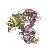
| ||||||||
| Unit cell |
| ||||||||
| Noncrystallographic symmetry (NCS) | NCS oper: (Code: given Matrix: (-0.987, -0.161, 0.009), Vector : |
-Components
| #1: Protein | Mass: 31192.322 Da / Num. of mol.: 2 Source method: isolated from a genetically manipulated source Details: FAD AND AMP COFACTORS ARE NONCOVALENTLY BOUND / Source: (gene. exp.) Paracoccus denitrificans (bacteria) / Plasmid: BLUESCRIPT / Production host: Escherichia coli (E. coli) / Strain (production host): DH5 ALPHA / References: UniProt: P38974#2: Protein | Mass: 26707.004 Da / Num. of mol.: 2 Source method: isolated from a genetically manipulated source Details: FAD AND AMP COFACTORS ARE NONCOVALENTLY BOUND / Source: (gene. exp.) Paracoccus denitrificans (bacteria) / Plasmid: BLUESCRIPT / Production host: Escherichia coli (E. coli) / Strain (production host): DH5 ALPHA / References: UniProt: P38975#3: Chemical | Flavin adenine dinucleotide  Mass: 785.550 Da / Num. of mol.: 2 / Source method: obtained synthetically / Formula: C27H33N9O15P2 / Comment: FAD*YM Mass: 785.550 Da / Num. of mol.: 2 / Source method: obtained synthetically / Formula: C27H33N9O15P2 / Comment: FAD*YM#4: Chemical | Adenosine monophosphate  Mass: 347.221 Da / Num. of mol.: 2 / Source method: obtained synthetically / Formula: C10H14N5O7P / Comment: AMP*YM Mass: 347.221 Da / Num. of mol.: 2 / Source method: obtained synthetically / Formula: C10H14N5O7P / Comment: AMP*YM#5: Water | ChemComp-HOH / | Water Mass: 18.015 Da / Num. of mol.: 101 / Source method: isolated from a natural source / Formula: H2O Mass: 18.015 Da / Num. of mol.: 101 / Source method: isolated from a natural source / Formula: H2O |
|---|
-Experimental details
-Experiment
| Experiment | Method: X-RAY DIFFRACTION / Number of used crystals: 1 |
|---|
- Sample preparation
| Crystal | Density Matthews: 2.33 Å3/Da / Density % sol: 49 % | ||||||||||||||||||||||||||||||||||||
|---|---|---|---|---|---|---|---|---|---|---|---|---|---|---|---|---|---|---|---|---|---|---|---|---|---|---|---|---|---|---|---|---|---|---|---|---|---|
| Crystal grow | pH: 5.8 Details: 18% PEG 8000, 25 MM KH2PO4, 100 UM FAD, AND 5 MM MGCL2, FINAL PH = 5.8. THE PROTEIN WAS MIXED WITH SOLUTION IN A 1:1 RATIO (PROTEIN WAS IN 10 MM TRIS, PH 7.4). | ||||||||||||||||||||||||||||||||||||
| Crystal | *PLUS | ||||||||||||||||||||||||||||||||||||
| Crystal grow | *PLUS Temperature: 19 ℃ / Method: vapor diffusion, hanging drop | ||||||||||||||||||||||||||||||||||||
| Components of the solutions | *PLUS
|
-Data collection
| Diffraction | Mean temperature: 297 K |
|---|---|
| Diffraction source | Source: ROTATING ANODE / Type: RIGAKU RU200 / Wavelength: 1.5418 |
| Detector | Type: RIGAKU RAXIS IIC / Detector: IMAGE PLATE / Date: Jan 1, 1995 |
| Radiation | Monochromator: GRAPHITE / Protocol: SINGLE WAVELENGTH / Monochromatic (M) / Laue (L): M / Scattering type: x-ray |
| Radiation wavelength | Wavelength: 1.5418 Å / Relative weight: 1 |
| Reflection | Resolution: 2.6→30 Å / Num. obs: 28666 / % possible obs: 87.8 % / Observed criterion σ(I): 0 / Redundancy: 4.2 % / Rmerge(I) obs: 0.054 / Rsym value: 0.054 |
| Reflection | *PLUS Num. measured all: 118452 |
- Processing
| Software |
| ||||||||||||||||||||||||||||||||||||||||||||||||||||||||||||
|---|---|---|---|---|---|---|---|---|---|---|---|---|---|---|---|---|---|---|---|---|---|---|---|---|---|---|---|---|---|---|---|---|---|---|---|---|---|---|---|---|---|---|---|---|---|---|---|---|---|---|---|---|---|---|---|---|---|---|---|---|---|
| Refinement | Method to determine structure: MOLECULAR REPLACEMENT Starting model: PDB ENTRY 1EFV Resolution: 2.6→12 Å / Rfactor Rfree error: 0.0061 / Cross valid method: THROUGHOUT / σ(F): 3
| ||||||||||||||||||||||||||||||||||||||||||||||||||||||||||||
| Displacement parameters | Biso mean: 26.2 Å2 | ||||||||||||||||||||||||||||||||||||||||||||||||||||||||||||
| Refinement step | Cycle: LAST / Resolution: 2.6→12 Å
| ||||||||||||||||||||||||||||||||||||||||||||||||||||||||||||
| Refine LS restraints |
| ||||||||||||||||||||||||||||||||||||||||||||||||||||||||||||
| LS refinement shell | Resolution: 2.6→2.75 Å / Total num. of bins used: 8
| ||||||||||||||||||||||||||||||||||||||||||||||||||||||||||||
| Xplor file | Serial no: 1 / Param file: PARAM19X.PRO / Topol file: TOPH19X.PRO | ||||||||||||||||||||||||||||||||||||||||||||||||||||||||||||
| Software | *PLUS Name: X-PLOR / Version: 3.851 / Classification: refinement | ||||||||||||||||||||||||||||||||||||||||||||||||||||||||||||
| Refinement | *PLUS Highest resolution: 2.6 Å / σ(F): 3 / % reflection Rfree: 7.5 % | ||||||||||||||||||||||||||||||||||||||||||||||||||||||||||||
| Solvent computation | *PLUS | ||||||||||||||||||||||||||||||||||||||||||||||||||||||||||||
| Displacement parameters | *PLUS Biso mean: 26.2 Å2 | ||||||||||||||||||||||||||||||||||||||||||||||||||||||||||||
| Refine LS restraints | *PLUS
| ||||||||||||||||||||||||||||||||||||||||||||||||||||||||||||
| LS refinement shell | *PLUS % reflection Rfree: 8.3 % / Rfactor Rwork: 0.259 |
