 Movie
Movie Controller

[English] 日本語
 Yorodumi
- PDB-1eez: Crystal Structure Determination of HLA-A2.1 Complexed to GP2 Pept... -
+ Open data

- Basic information
| Entry | Database: PDB / ID: 1eez | ||||||
|---|---|---|---|---|---|---|---|
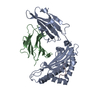
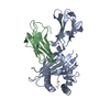
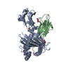
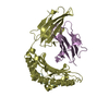
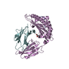
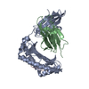
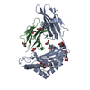
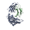
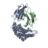
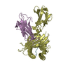
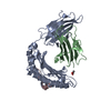
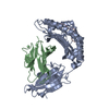
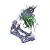
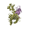
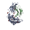
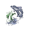
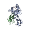
| Title | Crystal Structure Determination of HLA-A2.1 Complexed to GP2 Peptide Variant(I2L/V5L) | ||||||
 Components Components |
| ||||||
 Keywords Keywords |  IMMUNE SYSTEM / major histocompatibility complex / peptide binding IMMUNE SYSTEM / major histocompatibility complex / peptide binding | ||||||
| Function / homology |  Function and homology information: / : / retina homeostasis / T cell mediated cytotoxicity directed against tumor cell target / positive regulation of memory T cell activation / TAP complex binding / Golgi medial cisterna / positive regulation of CD8-positive, alpha-beta T cell activation / CD8-positive, alpha-beta T cell activation / positive regulation of CD8-positive, alpha-beta T cell proliferation ...: / : / retina homeostasis / T cell mediated cytotoxicity directed against tumor cell target / positive regulation of memory T cell activation / TAP complex binding / Golgi medial cisterna / positive regulation of CD8-positive, alpha-beta T cell activation / CD8-positive, alpha-beta T cell activation / positive regulation of CD8-positive, alpha-beta T cell proliferation / CD8 receptor binding / regulation of membrane depolarization / antigen processing and presentation of exogenous peptide antigen via MHC class I / endoplasmic reticulum exit site / antigen processing and presentation of endogenous peptide antigen via MHC class I via ER pathway, TAP-dependent / TAP binding / protection from natural killer cell mediated cytotoxicity / beta-2-microglobulin binding / detection of bacterium / T cell receptor binding / antigen processing and presentation of endogenous peptide antigen via MHC class I via ER pathway, TAP-independent / antigen processing and presentation of endogenous peptide antigen via MHC class Ib / positive regulation of ferrous iron binding / positive regulation of transferrin receptor binding / positive regulation of receptor binding / early endosome lumen / Nef mediated downregulation of MHC class I complex cell surface expression / DAP12 interactions / negative regulation of receptor binding / lumenal side of endoplasmic reticulum membrane / Endosomal/Vacuolar pathway / Antigen Presentation: Folding, assembly and peptide loading of class I MHC / cellular response to iron(III) ion / antigen processing and presentation of exogenous protein antigen via MHC class Ib, TAP-dependent / negative regulation of forebrain neuron differentiation / ER to Golgi transport vesicle membrane / regulation of erythrocyte differentiation / peptide antigen assembly with MHC class I protein complex / response to molecule of bacterial origin / regulation of iron ion transport / MHC class I peptide loading complex / HFE-transferrin receptor complex / T cell mediated cytotoxicity / cellular response to iron ion / antigen processing and presentation of endogenous peptide antigen via MHC class I / positive regulation of T cell cytokine production / MHC class I protein complex / multicellular organismal-level iron ion homeostasis / negative regulation of neurogenesis / peptide antigen assembly with MHC class II protein complex / positive regulation of receptor-mediated endocytosis / MHC class II protein complex / cellular response to nicotine / positive regulation of T cell mediated cytotoxicity / recycling endosome membrane / specific granule lumen / phagocytic vesicle membrane / peptide antigen binding / positive regulation of cellular senescence / antigen processing and presentation of exogenous peptide antigen via MHC class II / negative regulation of epithelial cell proliferation / Immunoregulatory interactions between a Lymphoid and a non-Lymphoid cell / Interferon gamma signaling / positive regulation of immune response / Modulation by Mtb of host immune system / positive regulation of type II interferon production / Interferon alpha/beta signaling / antimicrobial humoral immune response mediated by antimicrobial peptide / sensory perception of smell / positive regulation of T cell activation / positive regulation of protein binding / tertiary granule lumen / E3 ubiquitin ligases ubiquitinate target proteins / DAP12 signaling / negative regulation of neuron projection development / MHC class II protein complex binding / late endosome membrane / T cell differentiation in thymus / T cell receptor signaling pathway / ER-Phagosome pathway / iron ion transport / early endosome membrane / protein refolding / antibacterial humoral response / protein homotetramerization / intracellular iron ion homeostasis / cellular response to lipopolysaccharide / defense response to Gram-negative bacterium / amyloid fibril formation / learning or memory / defense response to Gram-positive bacterium / immune response / Amyloid fiber formation / lysosomal membrane / external side of plasma membrane / endoplasmic reticulum lumen / Golgi membrane / signaling receptor binding / focal adhesion / innate immune response Function and homology information: / : / retina homeostasis / T cell mediated cytotoxicity directed against tumor cell target / positive regulation of memory T cell activation / TAP complex binding / Golgi medial cisterna / positive regulation of CD8-positive, alpha-beta T cell activation / CD8-positive, alpha-beta T cell activation / positive regulation of CD8-positive, alpha-beta T cell proliferation ...: / : / retina homeostasis / T cell mediated cytotoxicity directed against tumor cell target / positive regulation of memory T cell activation / TAP complex binding / Golgi medial cisterna / positive regulation of CD8-positive, alpha-beta T cell activation / CD8-positive, alpha-beta T cell activation / positive regulation of CD8-positive, alpha-beta T cell proliferation / CD8 receptor binding / regulation of membrane depolarization / antigen processing and presentation of exogenous peptide antigen via MHC class I / endoplasmic reticulum exit site / antigen processing and presentation of endogenous peptide antigen via MHC class I via ER pathway, TAP-dependent / TAP binding / protection from natural killer cell mediated cytotoxicity / beta-2-microglobulin binding / detection of bacterium / T cell receptor binding / antigen processing and presentation of endogenous peptide antigen via MHC class I via ER pathway, TAP-independent / antigen processing and presentation of endogenous peptide antigen via MHC class Ib / positive regulation of ferrous iron binding / positive regulation of transferrin receptor binding / positive regulation of receptor binding / early endosome lumen / Nef mediated downregulation of MHC class I complex cell surface expression / DAP12 interactions / negative regulation of receptor binding / lumenal side of endoplasmic reticulum membrane / Endosomal/Vacuolar pathway / Antigen Presentation: Folding, assembly and peptide loading of class I MHC / cellular response to iron(III) ion / antigen processing and presentation of exogenous protein antigen via MHC class Ib, TAP-dependent / negative regulation of forebrain neuron differentiation / ER to Golgi transport vesicle membrane / regulation of erythrocyte differentiation / peptide antigen assembly with MHC class I protein complex / response to molecule of bacterial origin / regulation of iron ion transport / MHC class I peptide loading complex / HFE-transferrin receptor complex / T cell mediated cytotoxicity / cellular response to iron ion / antigen processing and presentation of endogenous peptide antigen via MHC class I / positive regulation of T cell cytokine production / MHC class I protein complex / multicellular organismal-level iron ion homeostasis / negative regulation of neurogenesis / peptide antigen assembly with MHC class II protein complex / positive regulation of receptor-mediated endocytosis / MHC class II protein complex / cellular response to nicotine / positive regulation of T cell mediated cytotoxicity / recycling endosome membrane / specific granule lumen / phagocytic vesicle membrane / peptide antigen binding / positive regulation of cellular senescence / antigen processing and presentation of exogenous peptide antigen via MHC class II / negative regulation of epithelial cell proliferation / Immunoregulatory interactions between a Lymphoid and a non-Lymphoid cell / Interferon gamma signaling / positive regulation of immune response / Modulation by Mtb of host immune system / positive regulation of type II interferon production / Interferon alpha/beta signaling / antimicrobial humoral immune response mediated by antimicrobial peptide / sensory perception of smell / positive regulation of T cell activation / positive regulation of protein binding / tertiary granule lumen / E3 ubiquitin ligases ubiquitinate target proteins / DAP12 signaling / negative regulation of neuron projection development / MHC class II protein complex binding / late endosome membrane / T cell differentiation in thymus / T cell receptor signaling pathway / ER-Phagosome pathway / iron ion transport / early endosome membrane / protein refolding / antibacterial humoral response / protein homotetramerization / intracellular iron ion homeostasis / cellular response to lipopolysaccharide / defense response to Gram-negative bacterium / amyloid fibril formation / learning or memory / defense response to Gram-positive bacterium / immune response / Amyloid fiber formation / lysosomal membrane / external side of plasma membrane / endoplasmic reticulum lumen / Golgi membrane / signaling receptor binding / focal adhesion / innate immune responseSimilarity search - Function | ||||||
| Biological species |  Homo sapiens (human) Homo sapiens (human) | ||||||
| Method | X-RAY DIFFRACTION / Resolution: 2.3 Å | ||||||
 Authors Authors | Sharma, A.K. / Kuhns, J.J. / Collins, E.J. | ||||||
 Citation Citation | Journal: J.Biol.Chem. / Year: 2001 Title: Class I major histocompatibility complex anchor substitutions alter the conformation of T cell receptor contacts. Authors: Sharma, A.K. / Kuhns, J.J. / Yan, S. / Friedline, R.H. / Long, B. / Tisch, R. / Collins, E.J. #1: Journal: J.Biol.Chem. / Year: 1999Title: Poor Binding of HER-2/neu Epitope (GP2) to HLA-A2.1 is Due to a Lack of Interactions with the Center of the Peptide Authors: Kuhns, J.J. / Batalia, M.A. / Yan, S. / Collins, E.J. | ||||||
| History |
|
- Structure visualization
| Structure viewer | Molecule: MolmilJmol/JSmol |
|---|
- Downloads & links
-Download
| PDBx/mmCIF format | 1eez.cif.gz | 159.1 KB | Display | PDBx/mmCIF format |
|---|---|---|---|---|
| PDB format | pdb1eez.ent.gz | 129.4 KB | Display | PDB format |
| PDBx/mmJSON format | 1eez.json.gz | Tree view | PDBx/mmJSON format | |
| Others |  Other downloads Other downloads |
-Validation report
| Arichive directory | https://data.pdbj.org/pub/pdb/validation_reports/ee/1eezftp://data.pdbj.org/pub/pdb/validation_reports/ee/1eez | HTTPS FTP |
|---|
-Related structure data


-Links
 PDBj
PDBj

- Assembly
| Deposited unit | 
| ||||||||
|---|---|---|---|---|---|---|---|---|---|
| 1 | 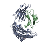
| ||||||||
| 2 | 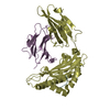
| ||||||||
| Unit cell |
| ||||||||
| Details | HLA_A2.1 heavy chain noncovalently associated to a beta-2-microglobulin subunit (light chain). Heavy chain is made of three sununits alpha1, alpha2 and alpha3. Peptide binds to the groove formed by the alpha1 and alpha2 domains |
-Components
| #1: Protein | Mass: 31854.203 Da / Num. of mol.: 2 / Fragment: RESIDUES 1-275 OF EXTRACELLULAR PORTION Source method: isolated from a genetically manipulated source Source: (gene. exp.) Homo sapiens (human) / Plasmid: PLM1 / Production host:  Escherichia coli (E. coli) / References: UniProt: P01892, UniProt: P04439*PLUS Escherichia coli (E. coli) / References: UniProt: P01892, UniProt: P04439*PLUS#2: Protein | Mass: 11879.356 Da / Num. of mol.: 2 Source method: isolated from a genetically manipulated source Source: (gene. exp.) Homo sapiens (human) / Plasmid: PLM1 / Production host: Escherichia coli (E. coli) / References: UniProt: P01884, UniProt: P61769*PLUS#3: Protein/peptide | Mass: 898.142 Da / Num. of mol.: 2 / Mutation: I2L,V5L / Source method: obtained synthetically Details: The peptide was synthesized at the peptide synthesis facility of University of North Carolina at Chapel Hill. #4: Water | ChemComp-HOH / | Water Mass: 18.015 Da / Num. of mol.: 35 / Source method: isolated from a natural source / Formula: H2O Mass: 18.015 Da / Num. of mol.: 35 / Source method: isolated from a natural source / Formula: H2OCompound details | HLA_A2.1 heavy chain noncovalently associated to a beta-2-microglobulin subunit (light chain). ...HLA_A2.1 heavy chain noncovalently associated to a beta-2-microglobulin subunit (light chain). Heavy chain is made of three subunits alpha1, alpha2 and alpha3. Peptide binds to the groove formed by the alpha1 and alpha2 domains. | |
|---|
-Experimental details
-Experiment
| Experiment | Method: X-RAY DIFFRACTION / Number of used crystals: 2 |
|---|
- Sample preparation
| Crystal | Density Matthews: 2.48 Å3/Da / Density % sol: 50.4 % | ||||||||||||||||||
|---|---|---|---|---|---|---|---|---|---|---|---|---|---|---|---|---|---|---|---|
| Crystal grow | Temperature: 298 K / Method: vapor diffusion, hanging drop / pH: 6.5 Details: 18% PEG8000, 25 mM MES, pH 6.5, VAPOR DIFFUSION, HANGING DROP, temperature 298K | ||||||||||||||||||
| Crystal grow | *PLUS Method: vapor diffusion, hanging drop | ||||||||||||||||||
| Components of the solutions | *PLUS
|
-Data collection
| Diffraction | Mean temperature: 100 K |
|---|---|
| Diffraction source | Source: ROTATING ANODE / Type: RIGAKU RU200 / Wavelength: 1.5418 |
| Detector | Type: RIGAKU RAXIS IIC / Detector: IMAGE PLATE / Date: Sep 30, 1999 |
| Radiation | Protocol: SINGLE WAVELENGTH / Monochromatic (M) / Laue (L): M / Scattering type: x-ray |
| Radiation wavelength | Wavelength: 1.5418 Å / Relative weight: 1 |
| Reflection | Resolution: 2.3→50 Å / Num. all: 114345 / Num. obs: 33403 / % possible obs: 87.7 % / Observed criterion σ(F): 0 / Observed criterion σ(I): 0 / Redundancy: 2 % / Biso Wilson estimate: 17.1 Å2 / Rmerge(I) obs: 0.095 / Net I/σ(I): 4.73 |
| Reflection shell | Resolution: 2.3→2.44 Å / Redundancy: 2 % / Rmerge(I) obs: 0.293 / Num. unique all: 5095 / % possible all: 84.8 |
| Reflection | *PLUS Lowest resolution: 50 Å / Num. measured all: 114345 |
| Reflection shell | *PLUS % possible obs: 84.8 % / Mean I/σ(I) obs: 2.4 |
- Processing
| Software |
| |||||||||||||||||||||||||
|---|---|---|---|---|---|---|---|---|---|---|---|---|---|---|---|---|---|---|---|---|---|---|---|---|---|---|
| Refinement | Resolution: 2.3→50 Å / Rfactor Rfree error: 0.007 / Data cutoff high absF: 974700.13 / Data cutoff low absF: 0 / Isotropic thermal model: RESTRAINED / Cross valid method: THROUGHOUT / σ(F): 0 / σ(I): 0 / Stereochemistry target values: Engh & Huber / Details: Maximum likelihood function
| |||||||||||||||||||||||||
| Solvent computation | Solvent model: FLAT MODEL / Bsol: 28.79 Å2 / ksol: 0.384 e/Å3 | |||||||||||||||||||||||||
| Displacement parameters | Biso mean: 21.3 Å2
| |||||||||||||||||||||||||
| Refine analyze |
| |||||||||||||||||||||||||
| Refinement step | Cycle: LAST / Resolution: 2.3→50 Å
| |||||||||||||||||||||||||
| Refine LS restraints |
| |||||||||||||||||||||||||
| LS refinement shell | Resolution: 2.3→2.44 Å / Rfactor Rfree error: 0.021 / Total num. of bins used: 6
| |||||||||||||||||||||||||
| Xplor file |
| |||||||||||||||||||||||||
| Refinement | *PLUS Highest resolution: 2.3 Å / Lowest resolution: 50 Å / % reflection Rfree: 5 % / Rfactor Rfree: 0.29 | |||||||||||||||||||||||||
| Solvent computation | *PLUS | |||||||||||||||||||||||||
| Displacement parameters | *PLUS | |||||||||||||||||||||||||
| Refine LS restraints | *PLUS
|
