 Movie
Movie Controller

[English] 日本語
 Yorodumi
- PDB-1eaw: Crystal structure of the MTSP1 (matriptase)-BPTI (aprotinin) complex -
+ Open data

- Basic information
| Entry | Database: PDB / ID: 1eaw | ||||||
|---|---|---|---|---|---|---|---|
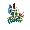
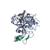
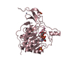
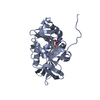
| Title | Crystal structure of the MTSP1 (matriptase)-BPTI (aprotinin) complex | ||||||
 Components Components |
| ||||||
 Keywords Keywords | HYDROLASE/INHIBITOR / COMPLEX (SERINE PROTEASE INHIBITOR) /  SERINE PROTEINASE / MATRIX DEGRADATION / INHIBITOR / GLYCOPROTE HYDROLASE / HYDROLASE-INHIBITOR complex SERINE PROTEINASE / MATRIX DEGRADATION / INHIBITOR / GLYCOPROTE HYDROLASE / HYDROLASE-INHIBITOR complex | ||||||
| Function / homology |  Function and homology informationmatriptase / epithelial cell morphogenesis involved in placental branching / acrosome reaction / trypsinogen activation / negative regulation of serine-type endopeptidase activity / Formation of the cornified envelope / sulfate binding / potassium channel inhibitor activity / negative regulation of platelet aggregation / zymogen binding ...matriptase / epithelial cell morphogenesis involved in placental branching / acrosome reaction / trypsinogen activation / negative regulation of serine-type endopeptidase activity / Formation of the cornified envelope / sulfate binding / potassium channel inhibitor activity / negative regulation of platelet aggregation / zymogen binding / molecular function inhibitor activity / negative regulation of thrombin-activated receptor signaling pathway / keratinocyte differentiation / serine protease inhibitor complex / serine-type peptidase activity / neural tube closure / protein catabolic process / serine-type endopeptidase inhibitor activity / basolateral plasma membrane / protease binding / external side of plasma membrane / serine-type endopeptidase activity / calcium ion binding / proteolysis / extracellular space / plasma membrane Function and homology informationmatriptase / epithelial cell morphogenesis involved in placental branching / acrosome reaction / trypsinogen activation / negative regulation of serine-type endopeptidase activity / Formation of the cornified envelope / sulfate binding / potassium channel inhibitor activity / negative regulation of platelet aggregation / zymogen binding ...matriptase / epithelial cell morphogenesis involved in placental branching / acrosome reaction / trypsinogen activation / negative regulation of serine-type endopeptidase activity / Formation of the cornified envelope / sulfate binding / potassium channel inhibitor activity / negative regulation of platelet aggregation / zymogen binding / molecular function inhibitor activity / negative regulation of thrombin-activated receptor signaling pathway / keratinocyte differentiation / serine protease inhibitor complex / serine-type peptidase activity / neural tube closure / protein catabolic process / serine-type endopeptidase inhibitor activity / basolateral plasma membrane / protease binding / external side of plasma membrane / serine-type endopeptidase activity / calcium ion binding / proteolysis / extracellular space / plasma membraneSimilarity search - Function | ||||||
| Biological species |  HOMO SAPIENS (human) HOMO SAPIENS (human) BOS TAURUS (cattle) BOS TAURUS (cattle) | ||||||
| Method | X-RAY DIFFRACTION / MOLECULAR REPLACEMENT / Resolution: 2.93 Å | ||||||
 Authors Authors | Friedrich, R. / Bode, W. | ||||||
 Citation Citation | Journal: J.Biol.Chem. / Year: 2002 Title: Catalytic Domain Structures of Mt-Sp1/Matriptase, a Matrix-Degrading Transmembrane Serine Proteinase. Authors: Friedrich, R. / Fuentes-Prior, P. / Ong, E. / Coombs, G. / Hunter, M. / Oehler, R. / Pierson, D. / Gonzalez, R. / Huber, R. / Bode, W. / Madison, E.L. | ||||||
| History |
| ||||||
| Remark 700 | SHEET DETERMINATION METHOD: DSSP THE SHEETS PRESENTED AS "AA" AND "CA" IN EACH CHAIN ON SHEET ... SHEET DETERMINATION METHOD: DSSP THE SHEETS PRESENTED AS "AA" AND "CA" IN EACH CHAIN ON SHEET RECORDS BELOW IS ACTUALLY AN 7-STRANDED BARREL THIS IS REPRESENTED BY A 8-STRANDED SHEET IN WHICH THE FIRST AND LAST STRANDS ARE IDENTICAL. THE SHEETS PRESENTED AS "AB" AND "CB" IN EACH CHAIN ON SHEET RECORDS BELOW IS ACTUALLY AN 6-STRANDED BARREL THIS IS REPRESENTED BY A 7-STRANDED SHEET IN WHICH THE FIRST AND LAST STRANDS ARE IDENTICAL. |
- Structure visualization
| Structure viewer | Molecule: MolmilJmol/JSmol |
|---|
- Downloads & links
-Download
| PDBx/mmCIF format | 1eaw.cif.gz | 125.8 KB | Display | PDBx/mmCIF format |
|---|---|---|---|---|
| PDB format | pdb1eaw.ent.gz | 97.6 KB | Display | PDB format |
| PDBx/mmJSON format | 1eaw.json.gz | Tree view | PDBx/mmJSON format | |
| Others |  Other downloads Other downloads |
-Validation report
| Arichive directory | https://data.pdbj.org/pub/pdb/validation_reports/ea/1eawftp://data.pdbj.org/pub/pdb/validation_reports/ea/1eaw | HTTPS FTP |
|---|
-Related structure data
| Related structure data | 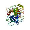 1eaxC  4htcS C: citing same article ( S: Starting model for refinement |
|---|---|
| Similar structure data |


-Links
 PDBj
PDBj


- Assembly
| Deposited unit | 
| ||||||||
|---|---|---|---|---|---|---|---|---|---|
| 1 | 
| ||||||||
| 2 | 
| ||||||||
| Unit cell |
|
-Components
| #1: Protein | Mass: 26463.756 Da / Num. of mol.: 2 / Fragment: CATALYTIC RESIDUES 615-855 Source method: isolated from a genetically manipulated source Source: (gene. exp.) HOMO SAPIENS (human) / Production host:  SACCHAROMYCES CEREVISIAE (brewer's yeast) SACCHAROMYCES CEREVISIAE (brewer's yeast)References: UniProt: Q9Y5Y6, Hydrolases; Acting on peptide bonds (peptidases); Serine endopeptidases#2: Protein | Mass: 6527.568 Da / Num. of mol.: 2 Source method: isolated from a genetically manipulated source Source: (gene. exp.) BOS TAURUS (cattle) / Production host:  ESCHERICHIA COLI (E. coli) / References: UniProt: P00974 ESCHERICHIA COLI (E. coli) / References: UniProt: P00974#3: Water | ChemComp-HOH / | Water Mass: 18.015 Da / Num. of mol.: 80 / Source method: isolated from a natural source / Formula: H2O Mass: 18.015 Da / Num. of mol.: 80 / Source method: isolated from a natural source / Formula: H2O |
|---|
-Experimental details
-Experiment
| Experiment | Method: X-RAY DIFFRACTION / Number of used crystals: 1 |
|---|
- Sample preparation
| Crystal | Density Matthews: 2.5 Å3/Da / Density % sol: 50 % / Description: 4HTC | ||||||||||||||||||||||||||||
|---|---|---|---|---|---|---|---|---|---|---|---|---|---|---|---|---|---|---|---|---|---|---|---|---|---|---|---|---|---|
| Crystal grow | pH: 8 / Details: pH 8.00 | ||||||||||||||||||||||||||||
| Crystal grow | *PLUS Temperature: 18 ℃ / Method: vapor diffusion, hanging drop / pH: 6.5 | ||||||||||||||||||||||||||||
| Components of the solutions | *PLUS
|
-Data collection
| Diffraction | Mean temperature: 100 K |
|---|---|
| Diffraction source | Source: ROTATING ANODE / Wavelength: 1.5418 |
| Detector | Type: MARRESEARCH / Detector: IMAGE PLATE / Date: Aug 15, 2000 |
| Radiation | Protocol: SINGLE WAVELENGTH / Monochromatic (M) / Laue (L): M / Scattering type: x-ray |
| Radiation wavelength | Wavelength: 1.5418 Å / Relative weight: 1 |
| Reflection | Resolution: 2.93→12 Å / Num. obs: 12200 / % possible obs: 94.4 % / Observed criterion σ(I): 2 / Redundancy: 2 % / Biso Wilson estimate: -0.1 Å2 / Rmerge(I) obs: 0.12 / Net I/σ(I): 5.4 |
| Reflection shell | Resolution: 2.93→3.11 Å / % possible all: 93.1 |
| Reflection | *PLUS Lowest resolution: 12 Å / Rmerge(I) obs: 0.12 |
| Reflection shell | *PLUS % possible obs: 93.1 % / Rmerge(I) obs: 0.343 |
- Processing
| Software |
| ||||||||||||||||||||||||||||||||||||||||||||||||||||||||||||
|---|---|---|---|---|---|---|---|---|---|---|---|---|---|---|---|---|---|---|---|---|---|---|---|---|---|---|---|---|---|---|---|---|---|---|---|---|---|---|---|---|---|---|---|---|---|---|---|---|---|---|---|---|---|---|---|---|---|---|---|---|---|
| Refinement | Method to determine structure: MOLECULAR REPLACEMENT Starting model: PDB ENTRY 4HTC Resolution: 2.93→11.99 Å / Rfactor Rfree error: 0.009 / Data cutoff high absF: 722936.73 / Cross valid method: THROUGHOUT / σ(F): 2
| ||||||||||||||||||||||||||||||||||||||||||||||||||||||||||||
| Solvent computation | Solvent model: MODEL | ||||||||||||||||||||||||||||||||||||||||||||||||||||||||||||
| Displacement parameters | Biso mean: 14.4 Å2
| ||||||||||||||||||||||||||||||||||||||||||||||||||||||||||||
| Refinement step | Cycle: LAST / Resolution: 2.93→11.99 Å
| ||||||||||||||||||||||||||||||||||||||||||||||||||||||||||||
| Refine LS restraints |
| ||||||||||||||||||||||||||||||||||||||||||||||||||||||||||||
| LS refinement shell | Resolution: 2.93→3.11 Å / Rfactor Rfree error: 0.033 / Total num. of bins used: 6
| ||||||||||||||||||||||||||||||||||||||||||||||||||||||||||||
| Xplor file |
| ||||||||||||||||||||||||||||||||||||||||||||||||||||||||||||
| Software | *PLUS Name: CNS / Version: 1 / Classification: refinement | ||||||||||||||||||||||||||||||||||||||||||||||||||||||||||||
| Refinement | *PLUS Lowest resolution: 12 Å | ||||||||||||||||||||||||||||||||||||||||||||||||||||||||||||
| Solvent computation | *PLUS | ||||||||||||||||||||||||||||||||||||||||||||||||||||||||||||
| Displacement parameters | *PLUS | ||||||||||||||||||||||||||||||||||||||||||||||||||||||||||||
| Refine LS restraints | *PLUS
| ||||||||||||||||||||||||||||||||||||||||||||||||||||||||||||
| LS refinement shell | *PLUS Rfactor Rwork: 0.3 |
