 Movie
Movie Controller


+ Open data

- Basic information
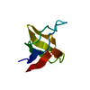
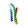
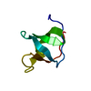
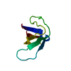
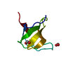
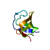
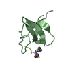
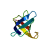
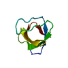
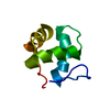
| Entry | Database: PDB / ID: 1e7o | ||||||
|---|---|---|---|---|---|---|---|
| Title | A-SPECTRIN SH3 DOMAIN A11V, V23L, M25V, V44I, V58L MUTATIONS | ||||||
 Components Components | SPECTRIN ALPHA CHAIN | ||||||
 Keywords Keywords |  STRUCTURAL PROTEIN / SH3-DOMAIN / CYTOSKELETON / CALMODULIN-BINDING / ACTIN-BINDING STRUCTURAL PROTEIN / SH3-DOMAIN / CYTOSKELETON / CALMODULIN-BINDING / ACTIN-BINDING | ||||||
| Function / homology |  Function and homology information Function and homology informationactin filament capping / costamere / cortical actin cytoskeleton / cell projection / actin filament binding / cell junction / actin cytoskeleton organization / calmodulin binding / calcium ion binding / plasma membraneSimilarity search - Function | ||||||
| Biological species |  GALLUS GALLUS (chicken) GALLUS GALLUS (chicken) | ||||||
| Method | X-RAY DIFFRACTION / MOLECULAR REPLACEMENT / Resolution: 3.2 Å | ||||||
 Authors Authors | Vega, M.C. / Serrano, L. | ||||||
 Citation Citation | Journal: J.Mol.Biol. / Year: 2003 Title: A Thermodynamic and Kinetic Analysis of the Folding Pathway of an SH3 Domain Entropically Stabilised by a Redesigned Hydrophobic Core Authors: Cobos, E.S. / Filimonov, V.V. / Vega, M.C. / Mateo, P.L. / Serrano, L. / Martinez, J.C. #1: Journal: Nature / Year: 1992Title: Crystal Structure of a Src-Homology 3 (SH3) Domain Authors: Musacchio, A. / Noble, M. / Pauptit, R. / Wierenga, R. / Saraste, M. | ||||||
| History |
| ||||||
| Remark 650 | HELIX DETERMINATION METHOD: AUTHOR PROVIDED. | ||||||
| Remark 700 | SHEET DETERMINATION METHOD: AUTHOR PROVIDED. |
- Structure visualization
| Structure viewer | Molecule: MolmilJmol/JSmol |
|---|
- Downloads & links
-Download
| PDBx/mmCIF format | 1e7o.cif.gz | 25.3 KB | Display | PDBx/mmCIF format |
|---|---|---|---|---|
| PDB format | pdb1e7o.ent.gz | 15.3 KB | Display | PDB format |
| PDBx/mmJSON format | 1e7o.json.gz | Tree view | PDBx/mmJSON format | |
| Others |  Other downloads Other downloads |
-Validation report
| Arichive directory | https://data.pdbj.org/pub/pdb/validation_reports/e7/1e7oftp://data.pdbj.org/pub/pdb/validation_reports/e7/1e7o | HTTPS FTP |
|---|
-Related structure data
| Related structure data | 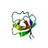 1shgS S: Starting model for refinement |
|---|---|
| Similar structure data |


-Links
 PDBj
PDBj- Assembly
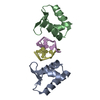
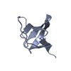
| Deposited unit | 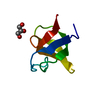
| ||||||||
|---|---|---|---|---|---|---|---|---|---|
| 1 |
| ||||||||
| Unit cell |
|
-Components
| #1: Protein | Mass: 7267.313 Da / Num. of mol.: 1 / Fragment: SH3-DOMAIN, RESIDUES 965-1025 / Mutation: YES Source method: isolated from a genetically manipulated source Source: (gene. exp.) GALLUS GALLUS (chicken) / Plasmid: PBR322 / Production host:  ESCHERICHIA COLI (E. coli) / Strain (production host): BL21(DE3) / References: UniProt: P07751 ESCHERICHIA COLI (E. coli) / Strain (production host): BL21(DE3) / References: UniProt: P07751 |
|---|---|
| #2: Chemical | ChemComp-GOL / Glycerol  Mass: 92.094 Da / Num. of mol.: 1 / Source method: obtained synthetically / Formula: C3H8O3 Mass: 92.094 Da / Num. of mol.: 1 / Source method: obtained synthetically / Formula: C3H8O3 |
| #3: Water | ChemComp-HOH / Water Mass: 18.015 Da / Num. of mol.: 20 / Source method: isolated from a natural source / Formula: H2O Mass: 18.015 Da / Num. of mol.: 20 / Source method: isolated from a natural source / Formula: H2O |
| Compound details | CHAIN A ENGINEERED MUTATION ALA 974 VAL, VAL 986 LEU, MET 988 VAL, VAL 1007 ILE AND VAL 1021 LEU ...CHAIN A ENGINEERED |
-Experimental details
-Experiment
| Experiment | Method: X-RAY DIFFRACTION / Number of used crystals: 1 |
|---|
- Sample preparation
| Crystal | Density Matthews: 2.25 Å3/Da / Density % sol: 45.5 % | ||||||||||||
|---|---|---|---|---|---|---|---|---|---|---|---|---|---|
| Crystal grow | pH: 6 Details: PROTEIN WAS CRYSTALLIZED FROM 1.1 M AMMONIUM SULPHATE, 90MM SODIUM CITRATE/CITRIC ACID, PH=6.0, 90 MM BIS-TRIS PROPANE, 0.9 MM EDTA, 0.9 MM DTT, 0.9 MM SODIUM AZIDE, pH 6.00 | ||||||||||||
| Crystal grow | *PLUS Temperature: 20 ℃ / Method: vapor diffusion | ||||||||||||
| Components of the solutions | *PLUS
|
-Data collection
| Diffraction | Mean temperature: 100 K |
|---|---|
| Diffraction source | Source: ROTATING ANODE / Type: MACSCIENCE M18X / Wavelength: 1.5418 |
| Detector | Type: MARRESEARCH / Detector: IMAGE PLATE / Date: May 15, 2000 / Details: MIRROR |
| Radiation | Protocol: SINGLE WAVELENGTH / Monochromatic (M) / Laue (L): M / Scattering type: x-ray |
| Radiation wavelength | Wavelength: 1.5418 Å / Relative weight: 1 |
| Reflection | Resolution: 3.2→12 Å / Num. obs: 1003 / % possible obs: 82.9 % / Observed criterion σ(I): 2 / Redundancy: 2.3 % / Rmerge(I) obs: 0.134 |
| Reflection shell | Resolution: 3.2→3.5 Å / Rmerge(I) obs: 0.138 / % possible all: 77.1 |
| Reflection | *PLUS Highest resolution: 3 Å / Lowest resolution: 10 Å / Num. obs: 1172 |
- Processing
| Software |
| ||||||||||||||||||||||||||||||||||||||||||||||||||||||||||||
|---|---|---|---|---|---|---|---|---|---|---|---|---|---|---|---|---|---|---|---|---|---|---|---|---|---|---|---|---|---|---|---|---|---|---|---|---|---|---|---|---|---|---|---|---|---|---|---|---|---|---|---|---|---|---|---|---|---|---|---|---|---|
| Refinement | Method to determine structure: MOLECULAR REPLACEMENT Starting model: PDB ENTRY 1SHG Resolution: 3.2→8 Å / Cross valid method: THROUGHOUT / σ(F): 2 Details: THE FIRST RESIDUE IN N-TERMINAL WAS NOT SEEN IN THE DENSITY MAPS
| ||||||||||||||||||||||||||||||||||||||||||||||||||||||||||||
| Refinement step | Cycle: LAST / Resolution: 3.2→8 Å
| ||||||||||||||||||||||||||||||||||||||||||||||||||||||||||||
| Refine LS restraints |
| ||||||||||||||||||||||||||||||||||||||||||||||||||||||||||||
| LS refinement shell | Resolution: 3.2→3.34 Å | ||||||||||||||||||||||||||||||||||||||||||||||||||||||||||||
| Xplor file |
| ||||||||||||||||||||||||||||||||||||||||||||||||||||||||||||
| Refinement | *PLUS Highest resolution: 3 Å / Lowest resolution: 10 Å / Num. reflection obs: 1172 / Rfactor Rfree: 0.275 / Rfactor Rwork: 0.2334 | ||||||||||||||||||||||||||||||||||||||||||||||||||||||||||||
| Solvent computation | *PLUS | ||||||||||||||||||||||||||||||||||||||||||||||||||||||||||||
| Displacement parameters | *PLUS | ||||||||||||||||||||||||||||||||||||||||||||||||||||||||||||
| Refine LS restraints | *PLUS
|
