 Movie
Movie Controller


+ Open data

- Basic information
| Entry | Database: PDB / ID: 1.0E+58 | ||||||
|---|---|---|---|---|---|---|---|
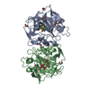
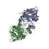
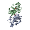
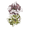
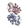
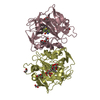
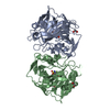
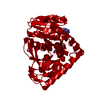
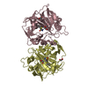
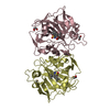
| Title | E.coli cofactor-dependent phosphoglycerate mutase | ||||||
 Components Components | PHOSPHOGLYCERATE MUTASE | ||||||
 Keywords Keywords | ISOMERASE / PHOSPHOHISTIDINE / GLYCOLYSIS AND GLUCONEOGENESIS / PHOSPHOGLYCERATE MUTASE | ||||||
| Function / homology |  Function and homology information Function and homology information2,3-bisphosphoglycerate-dependent phosphoglycerate mutase activity / phosphoglycerate mutase (2,3-diphosphoglycerate-dependent) / canonical glycolysis / guanosine tetraphosphate binding / gluconeogenesis / protein homodimerization activity / ATP binding / cytosol / cytoplasmSimilarity search - Function | ||||||
| Biological species |  ESCHERICHIA COLI (E. coli) ESCHERICHIA COLI (E. coli) | ||||||
| Method | X-RAY DIFFRACTION / SYNCHROTRON / MOLECULAR REPLACEMENT / Resolution: 1.25 Å | ||||||
 Authors Authors | Bond, C.S. / Hunter, W.N. | ||||||
 Citation Citation | Journal: J.Biol.Chem. / Year: 2001 Title: High Resolution Structure of the Phosphohistidine-Activated Form of Escherichia Coli Cofactor-Dependent Phosphoglycerate Mutase. Authors: Bond, C.S. / White, M.F. / Hunter, W.N. | ||||||
| History |
|
- Structure visualization
| Structure viewer | Molecule: MolmilJmol/JSmol |
|---|
- Downloads & links
-Download
| PDBx/mmCIF format | 1e58.cif.gz | 129.5 KB | Display | PDBx/mmCIF format |
|---|---|---|---|---|
| PDB format | pdb1e58.ent.gz | 106 KB | Display | PDB format |
| PDBx/mmJSON format | 1e58.json.gz | Tree view | PDBx/mmJSON format | |
| Others |  Other downloads Other downloads |
-Validation report
| Arichive directory | https://data.pdbj.org/pub/pdb/validation_reports/e5/1e58ftp://data.pdbj.org/pub/pdb/validation_reports/e5/1e58 | HTTPS FTP |
|---|
-Related structure data
| Related structure data | |
|---|---|
| Similar structure data |

-Links
 PDBj
PDBj


- Assembly
| Deposited unit | 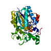
| ||||||||
|---|---|---|---|---|---|---|---|---|---|
| 1 | 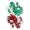
| ||||||||
| Unit cell |
|
-Components
| #1: Protein | Mass: 28544.164 Da / Num. of mol.: 1 Source method: isolated from a genetically manipulated source Source: (gene. exp.) ESCHERICHIA COLI (E. coli) / Strain: K12 / Gene: PGM1 / Plasmid: PET3A / Production host: ESCHERICHIA COLI (E. coli) / Strain (production host): BL21(DE3) / References: UniProt: P62707, EC: 5.4.2.1 | ||||||
|---|---|---|---|---|---|---|---|
| #2: Chemical | Sulfate  Mass: 96.063 Da / Num. of mol.: 2 / Source method: obtained synthetically / Formula: SO4 Mass: 96.063 Da / Num. of mol.: 2 / Source method: obtained synthetically / Formula: SO4#3: Chemical | ChemComp-CL / | Chloride  Mass: 35.453 Da / Num. of mol.: 1 / Source method: obtained synthetically / Formula: Cl Mass: 35.453 Da / Num. of mol.: 1 / Source method: obtained synthetically / Formula: Cl#4: Water | ChemComp-HOH / | Water Mass: 18.015 Da / Num. of mol.: 425 / Source method: isolated from a natural source / Formula: H2O Mass: 18.015 Da / Num. of mol.: 425 / Source method: isolated from a natural source / Formula: H2OCompound details | CATALYTIC ACTIVITY: 2-PHOSPHOGLYCERATE + 2,3-DIPHOSPHOGLYCERATE = 3-PHOSPHOGLYCERATE + 2,3- ...CATALYTIC ACTIVITY: 2-PHOSPHOGLY | |
-Experimental details
-Experiment
| Experiment | Method: X-RAY DIFFRACTION / Number of used crystals: 1 |
|---|
- Sample preparation
| Crystal | Density Matthews: 2.14 Å3/Da / Density % sol: 51 % | ||||||||||||||||||||||||||||||||||||||||||
|---|---|---|---|---|---|---|---|---|---|---|---|---|---|---|---|---|---|---|---|---|---|---|---|---|---|---|---|---|---|---|---|---|---|---|---|---|---|---|---|---|---|---|---|
| Crystal grow | pH: 8.5 Details: 100 MM TRIS-HCL (PH 8.0), 200 MM LI2SO4, 20% PEG 4000 | ||||||||||||||||||||||||||||||||||||||||||
| Crystal | *PLUS Density % sol: 50 % | ||||||||||||||||||||||||||||||||||||||||||
| Crystal grow | *PLUS pH: 8 / Method: vapor diffusion, hanging drop | ||||||||||||||||||||||||||||||||||||||||||
| Components of the solutions | *PLUS
|
-Data collection
| Diffraction | Mean temperature: 105 K |
|---|---|
| Diffraction source | Source: SYNCHROTRON / Site: SRS  / Beamline: PX9.6 / Wavelength: 0.89 / Beamline: PX9.6 / Wavelength: 0.89 |
| Detector | Date: Sep 15, 1999 |
| Radiation | Protocol: SINGLE WAVELENGTH / Monochromatic (M) / Laue (L): M / Scattering type: x-ray |
| Radiation wavelength | Wavelength: 0.89 Å / Relative weight: 1 |
| Reflection | Resolution: 1.25→30 Å / Num. obs: 67122 / % possible obs: 85.4 % / Redundancy: 2 % / Rsym value: 0.047 / Net I/σ(I): 211 |
| Reflection shell | Resolution: 1.25→1.26 Å / Redundancy: 2 % / Mean I/σ(I) obs: 11 / Rsym value: 0.071 / % possible all: 63 |
| Reflection | *PLUS Rmerge(I) obs: 0.047 |
| Reflection shell | *PLUS % possible obs: 63 % / Rmerge(I) obs: 0.071 / Mean I/σ(I) obs: 11.2 |
- Processing
| Software |
| |||||||||||||||||||||||||||||||||
|---|---|---|---|---|---|---|---|---|---|---|---|---|---|---|---|---|---|---|---|---|---|---|---|---|---|---|---|---|---|---|---|---|---|---|
| Refinement | Method to determine structure: MOLECULAR REPLACEMENT Starting model: NONE Resolution: 1.25→10 Å / Num. parameters: 22606 / Num. restraintsaints: 27457 / Cross valid method: FREE R-VALUE / σ(F): 0 / Stereochemistry target values: ENGH AND HUBER Details: ANISOTROPIC SCALING APPLIED BY THE METHOD OF PARKIN, MOEZZI & HOPE, J.APPL.CRYST.28 (1995)53-56 PHOSPHOHISTIDINE MODELLED AT 0.28 OCCUPANCY COUPLED TO A 0.72 OCCUPANCY HISTIDINE WITH THREE WATER MOLECULES
| |||||||||||||||||||||||||||||||||
| Solvent computation | Solvent model: MOEWS & KRETSINGER, J.MOL.BIOL.91(1973)201-2 | |||||||||||||||||||||||||||||||||
| Refine analyze | Num. disordered residues: 17 / Occupancy sum hydrogen: 1977 / Occupancy sum non hydrogen: 2434.16 | |||||||||||||||||||||||||||||||||
| Refinement step | Cycle: LAST / Resolution: 1.25→10 Å
| |||||||||||||||||||||||||||||||||
| Refine LS restraints |
| |||||||||||||||||||||||||||||||||
| Software | *PLUS Name: SHELXL-97 / Classification: refinement | |||||||||||||||||||||||||||||||||
| Refinement | *PLUS | |||||||||||||||||||||||||||||||||
| Solvent computation | *PLUS | |||||||||||||||||||||||||||||||||
| Displacement parameters | *PLUS Biso mean: 14 Å2 |
