 Movie
Movie Controller


+ Open data

- Basic information
| Entry | Database: PDB / ID: 1dxy | ||||||
|---|---|---|---|---|---|---|---|
| Title | STRUCTURE OF D-2-HYDROXYISOCAPROATE DEHYDROGENASE | ||||||
 Components Components | D-2-HYDROXYISOCAPROATE DEHYDROGENASE | ||||||
 Keywords Keywords |  OXIDOREDUCTASE / D-2-HYDROXYCARBOXYLATE DEHYDROGENASE / D-LACTATE DEHYDROGENASE OXIDOREDUCTASE / D-2-HYDROXYCARBOXYLATE DEHYDROGENASE / D-LACTATE DEHYDROGENASE | ||||||
| Function / homology |  Function and homology informationOxidoreductases; Acting on the CH-OH group of donors; With NAD+ or NADP+ as acceptor / oxidoreductase activity, acting on the CH-OH group of donors, NAD or NADP as acceptor / NAD binding Function and homology informationOxidoreductases; Acting on the CH-OH group of donors; With NAD+ or NADP+ as acceptor / oxidoreductase activity, acting on the CH-OH group of donors, NAD or NADP as acceptor / NAD bindingSimilarity search - Function | ||||||
| Biological species |  Lactobacillus casei (bacteria) Lactobacillus casei (bacteria) | ||||||
| Method | X-RAY DIFFRACTION / SYNCHROTRON / MOLECULAR REPLACEMENT / Resolution: 1.86 Å | ||||||
 Authors Authors | Dengler, U. / Niefind, K. / Kiess, M. / Schomburg, D. | ||||||
 Citation Citation | Journal: J.Mol.Biol. / Year: 1997 Title: Crystal structure of a ternary complex of D-2-hydroxyisocaproate dehydrogenase from Lactobacillus casei, NAD+ and 2-oxoisocaproate at 1.9 A resolution. Authors: Dengler, U. / Niefind, K. / Kiess, M. / Schomburg, D. #1: Journal: J.Mol.Biol. / Year: 1994Title: Crystallization and Preliminary Characterization of Crystals of D-2-Hydroxyisocaproate Dehydrogenase from Lactobacillus Casei Authors: Niefind, K. / Hecht, H.J. / Schomburg, D. #2: Journal: Thesis / Year: 1993Title: Roentgenkristallographische Untersuchungen an Drei Mikrobiellen Enzymen: D-2-Hydroxyisocaproat-Dehydrogenase Aus Lactobacillus Casei L-2-Hydroxyisocaproat-Dehydrogenase Aus Lactobacillus ...Title: Roentgenkristallographische Untersuchungen an Drei Mikrobiellen Enzymen: D-2-Hydroxyisocaproat-Dehydrogenase Aus Lactobacillus Casei L-2-Hydroxyisocaproat-Dehydrogenase Aus Lactobacillus Confusus Alkalische Protease Aus Bacillus Alcalophilus/Variante Q59R Authors: Niefind, K. #3: Journal: ENZYME.MICROB.TECHNOL. / Year: 1992Title: Potential of R-2-Hydroxyisocaproate Dehydrogenase from Lactobacillus Casei for Stereospecific Reductions Authors: Kallwass, H.K.W. #4: Journal: Gene / Year: 1989Title: Cloning, Sequencing and Expression in Escherichia Coli of the D-2-Hydroxyisocaproate Dehydrogenase Gene of Lactobacillus Casei Authors: Lerch, H.P. / Blocker, H. / Kallwass, H. / Hoppe, J. / Tsai, H. / Collins, J. #5: Journal: Fems Microbiol.Lett. / Year: 1987Title: Crystallization and Molecular Properties of D-2-Hydroxyisocaproate Dehydrogenase from Lactobacillus Casei Authors: Kallwass, H. / Tsai, H. / Schuette, H. #6: Journal: Appl.Microbiol.Biotechnol. / Year: 1985Title: D-2-Hydroxyisocaproate Dehydrogenase from Lactobacillus Casei. A New Enzyme Suitable for Stereospecific Reduction of 2-Ketocarboxylic Acids Authors: Hummel, W. / Schuette, H. / Kula, M.R. | ||||||
| History |
|
- Structure visualization
| Structure viewer | Molecule: MolmilJmol/JSmol |
|---|
- Downloads & links
-Download
| PDBx/mmCIF format | 1dxy.cif.gz | 80.6 KB | Display | PDBx/mmCIF format |
|---|---|---|---|---|
| PDB format | pdb1dxy.ent.gz | 63.5 KB | Display | PDB format |
| PDBx/mmJSON format | 1dxy.json.gz | Tree view | PDBx/mmJSON format | |
| Others |  Other downloads Other downloads |
-Validation report
| Arichive directory | https://data.pdbj.org/pub/pdb/validation_reports/dx/1dxyftp://data.pdbj.org/pub/pdb/validation_reports/dx/1dxy | HTTPS FTP |
|---|
-Related structure data
| Related structure data |  1gdhS S: Starting model for refinement |
|---|---|
| Similar structure data |


-Links
 PDBj
PDBj- Assembly
| Deposited unit | 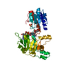
| |||||||||
|---|---|---|---|---|---|---|---|---|---|---|
| 1 | 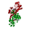
| |||||||||
| 2 | x 6
| |||||||||
| Unit cell |
| |||||||||
| Components on special symmetry positions |
| |||||||||
| Details | D-HICDH IS A DIMER OF IDENTICAL SUBUNITS, WHICH OCCUPIES A SPECIAL POSITION IN THE INVESTIGATED CRYSTALS. |
-Components
| #1: Protein | Mass: 36929.840 Da / Num. of mol.: 1 Source method: isolated from a genetically manipulated source Source: (gene. exp.) Lactobacillus casei (bacteria) / Gene: POTENTIAL / Variant: SSP. PSEUDOPLANTARUM / Plasmid: PJLA601 / Production host: Escherichia coli (E. coli)References: UniProt: P17584, Oxidoreductases; Acting on the CH-OH group of donors; With NAD+ or NADP+ as acceptor | ||||||||||
|---|---|---|---|---|---|---|---|---|---|---|---|
| #2: Chemical | Sulfate  Mass: 96.063 Da / Num. of mol.: 2 / Source method: obtained synthetically / Formula: SO4 Mass: 96.063 Da / Num. of mol.: 2 / Source method: obtained synthetically / Formula: SO4#3: Chemical | ChemComp-NAD / | Nicotinamide adenine dinucleotide  Mass: 663.425 Da / Num. of mol.: 1 / Source method: obtained synthetically / Formula: C21H27N7O14P2 / Comment: NAD*YM Mass: 663.425 Da / Num. of mol.: 1 / Source method: obtained synthetically / Formula: C21H27N7O14P2 / Comment: NAD*YM#4: Chemical | ChemComp-COI / | Alpha-Ketoisocaproic acid  Mass: 130.142 Da / Num. of mol.: 1 / Source method: obtained synthetically / Formula: C6H10O3 Mass: 130.142 Da / Num. of mol.: 1 / Source method: obtained synthetically / Formula: C6H10O3#5: Water | ChemComp-HOH / | Water Mass: 18.015 Da / Num. of mol.: 182 / Source method: isolated from a natural source / Formula: H2O Mass: 18.015 Da / Num. of mol.: 182 / Source method: isolated from a natural source / Formula: H2ONonpolymer details | 2-OXOISOCAPROATE (OIC 337) AND A SULFATE ION (SO4 338) ARE MUTUALLY EXCLUSIVELY BOUND IN THE ACTIVE ...2-OXOISOCAPR | Sequence details | A CONTACT IS OBSERVED BETWEEN THE SIDE CHAINS OF ASP 266 AND ASP 278. CHEMICAL PROTEIN SEQUENCING ...A CONTACT IS OBSERVED BETWEEN THE SIDE CHAINS OF ASP 266 AND ASP 278. CHEMICAL PROTEIN SEQUENCING | |
-Experimental details
-Experiment
| Experiment | Method: X-RAY DIFFRACTION / Number of used crystals: 3 |
|---|
- Sample preparation
| Crystal | Density Matthews: 4.4 Å3/Da / Density % sol: 72 % | ||||||||||||||||||||||||||||||||||||||||
|---|---|---|---|---|---|---|---|---|---|---|---|---|---|---|---|---|---|---|---|---|---|---|---|---|---|---|---|---|---|---|---|---|---|---|---|---|---|---|---|---|---|
| Crystal grow | Temperature: 277 K / pH: 7 Details: 1.9 M AMMONIUM SULFATE, 50 MM CITRATE/PHOSPHATE BUFFER, 30 MM NAD+, 60 MM 4-METHYL-2-OXOPENTANOATE, PH 7.0, 277 K; ENZYME CONCENTRATION 10 MG/ML. | ||||||||||||||||||||||||||||||||||||||||
| Crystal grow | *PLUS Temperature: 4 ℃ / Method: vapor diffusion, hanging drop / Details: Niefind, K., (1994) J.Mol.Biol., 240, 400. | ||||||||||||||||||||||||||||||||||||||||
| Components of the solutions | *PLUS
|
-Data collection
| Diffraction | Mean temperature: 277 K |
|---|---|
| Diffraction source | Source: SYNCHROTRON / Site: MPG/DESY, HAMBURG  / Beamline: BW6 / Wavelength: 1.1 / Beamline: BW6 / Wavelength: 1.1 |
| Detector | Type: MAR scanner 300 mm plate / Detector: IMAGE PLATE / Date: Aug 1, 1994 / Details: MIRROR OPTICS |
| Radiation | Monochromator: SI(111) / Monochromatic (M) / Laue (L): M / Scattering type: x-ray |
| Radiation wavelength | Wavelength: 1.1 Å / Relative weight: 1 |
| Reflection | Resolution: 1.86→12.1 Å / Num. obs: 53838 / % possible obs: 96.5 % / Observed criterion σ(I): 0 / Redundancy: 5.8 % / Rsym value: 0.089 / Net I/σ(I): 22.2 |
| Reflection shell | Resolution: 1.86→2 Å / Redundancy: 4.4 % / Mean I/σ(I) obs: 2.8 / Rsym value: 0.48 / % possible all: 86.9 |
| Reflection | *PLUS Num. measured all: 314570 / Rmerge(I) obs: 0.089 |
| Reflection shell | *PLUS % possible obs: 86.9 % / Rmerge(I) obs: 0.48 |
- Processing
| Software |
| ||||||||||||||||||||||||||||||||||||||||||||||||||||||||||||
|---|---|---|---|---|---|---|---|---|---|---|---|---|---|---|---|---|---|---|---|---|---|---|---|---|---|---|---|---|---|---|---|---|---|---|---|---|---|---|---|---|---|---|---|---|---|---|---|---|---|---|---|---|---|---|---|---|---|---|---|---|---|
| Refinement | Method to determine structure: MOLECULAR REPLACEMENT Starting model: BACKBONE AND C-BETA ATOMS OF D-GLYCERATE DEHYDROGENASE (PDB ENTRY 1GDH) Resolution: 1.86→12.1 Å / σ(F): 0
| ||||||||||||||||||||||||||||||||||||||||||||||||||||||||||||
| Displacement parameters | Biso mean: 31.02 Å2 | ||||||||||||||||||||||||||||||||||||||||||||||||||||||||||||
| Refine analyze | Luzzati coordinate error obs: 0.23 Å / Luzzati sigma a obs: 0.28 Å | ||||||||||||||||||||||||||||||||||||||||||||||||||||||||||||
| Refinement step | Cycle: LAST / Resolution: 1.86→12.1 Å
| ||||||||||||||||||||||||||||||||||||||||||||||||||||||||||||
| Refine LS restraints |
| ||||||||||||||||||||||||||||||||||||||||||||||||||||||||||||
| Software | *PLUS Name: X-PLOR / Version: 3.1 / Classification: refinement | ||||||||||||||||||||||||||||||||||||||||||||||||||||||||||||
| Refine LS restraints | *PLUS
|
