 Movie
Movie Controller

[English] 日本語
 Yorodumi
- PDB-1dxr: Photosynthetic reaction center from Rhodopseudomonas viridis - Hi... -
+ Open data

- Basic information
| Entry | Database: PDB / ID: 1dxr | ||||||
|---|---|---|---|---|---|---|---|
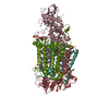
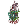
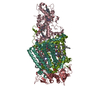
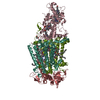
| Title | Photosynthetic reaction center from Rhodopseudomonas viridis - His L168 Phe mutant (terbutryn complex) | ||||||
 Components Components | (PHOTOSYNTHETIC REACTION CENTER ... Photosynthetic reaction centre) x 4 Photosynthetic reaction centre) x 4 | ||||||
 Keywords Keywords | PHOTOSYNTHESIS / SECONDARY QUINONE (QB) / TRIAZINE INHIBITOR | ||||||
| Function / homology |  Function and homology information Function and homology informationplasma membrane-derived chromatophore membrane / plasma membrane light-harvesting complex / bacteriochlorophyll binding / photosynthesis, light reaction / electron transporter, transferring electrons within the cyclic electron transport pathway of photosynthesis activity / photosynthetic electron transport in photosystem II / photosynthesis / electron transfer activity / iron ion binding / heme binding / metal ion bindingSimilarity search - Function | ||||||
| Biological species |  RHODOPSEUDOMONAS VIRIDIS (bacteria) RHODOPSEUDOMONAS VIRIDIS (bacteria) | ||||||
| Method | X-RAY DIFFRACTION / SYNCHROTRON / MOLECULAR REPLACEMENT / Resolution: 2 Å | ||||||
 Authors Authors | Lancaster, C.R.D. / Bibikova, M. / Sabatino, P. / Oesterhelt, D. / Michel, H. | ||||||
 Citation Citation | Journal: J. Biol. Chem. / Year: 2000 Title: Structural basis of the drastically increased initial electron transfer rate in the reaction center from a Rhodopseudomonas viridis mutant described at 2.00-A resolution. Authors: Lancaster, C.R. / Bibikova, M.V. / Sabatino, P. / Oesterhelt, D. / Michel, H. #1: Journal: J.Mol.Biol. / Year: 1999Title: Refined Crystal Structures of Reaction Centres from Rhodopseudomonas Viridis in Complexes with the Herbicide Atrazine and Two Chiral Atrazine Derivatives Also Lead to a New Model of the Bound Carotenoid Authors: Lancaster, C.R. / Michel, H. #2: Journal: Biochim.Biophys.Acta / Year: 1998Title: Ubiquinone Reduction and Protonation in the Reaction Centre of Rhodopseudomonas Viridis: X-Ray Structures and Their Functional Implications Authors: Lancaster, C.R.D. #3: Journal: Structure / Year: 1997Title: The Coupling of Light-Induced Electron Transfer and Proton Uptake as Derived from Crystal Structures of Reaction Centres from Rhodopseudomonas Viridis Modified at the Binding Site of the Secondary Quinone, Qb Authors: Lancaster, C.R. / Michel, H. #4: Journal: J.Mol.Biol. / Year: 1995Title: Crystallographic Refinement at 2.3 A Resolution and Refined Model of the Photosynthetic Reaction Centre from Rhodopseudomonas Viridis Authors: Deisenhofer, J. / Epp, O. / Sinning, I. / Michel, H. #5: Journal: Science / Year: 1989 Title: The Photosynthetic Reaction Center from the Purple Bacterium Rhodopseudomonas Viridis Authors: Deisenhofer, J. / Michel, H. #6: Journal: Nature / Year: 1985 Title: Structure of the Protein Subunits in the Photosynthetic Reaction Centre of Rhodopseudomonas Viridis at 3 Angstroms Resolution Authors: Deisenhofer, J. / Epp, O. / Miki, K. / Huber, R. / Michel, H. #7: Journal: J.Mol.Biol. / Year: 1984 Title: X-Ray Structure Analysis of a Membrane Protein Complex. Electron Density Map at 3 A Resolution and a Model of the Chromophores of the Photosynthetic Reaction Center from Rhodopseudomonas Viridis Authors: Deisenhofer, J. / Epp, O. / Miki, K. / Huber, R. / Michel, H. #8: Journal: J.Mol.Biol. / Year: 1982 Title: Three-Dimensional Crystals of a Membrane Protein Complex. The Photosynthetic Reaction Centre from Rhodopseudomonas Viridis Authors: Michel, H. | ||||||
| History |
| ||||||
| Remark 650 | HELIX DETERMINATION METHOD: AUTHOR PROVIDED. | ||||||
| Remark 700 | SHEET DETERMINATION METHOD: AUTHOR PROVIDED. |
- Structure visualization
| Structure viewer | Molecule: MolmilJmol/JSmol |
|---|
- Downloads & links
-Download
| PDBx/mmCIF format | 1dxr.cif.gz | 290.3 KB | Display | PDBx/mmCIF format |
|---|---|---|---|---|
| PDB format | pdb1dxr.ent.gz | 227.7 KB | Display | PDB format |
| PDBx/mmJSON format | 1dxr.json.gz | Tree view | PDBx/mmJSON format | |
| Others |  Other downloads Other downloads |
-Validation report
| Arichive directory | https://data.pdbj.org/pub/pdb/validation_reports/dx/1dxrftp://data.pdbj.org/pub/pdb/validation_reports/dx/1dxr | HTTPS FTP |
|---|
-Related structure data
| Related structure data |  6prcS S: Starting model for refinement |
|---|---|
| Similar structure data |


-Links
 PDBj
PDBj


- Assembly
| Deposited unit | 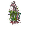
| ||||||||
|---|---|---|---|---|---|---|---|---|---|
| 1 |
| ||||||||
| Unit cell |
|
-Components
-PHOTOSYNTHETIC REACTION CENTER ... , 4 types, 4 molecules CHLM
| #1: Protein | Mass: 37450.801 Da / Num. of mol.: 1 / Source method: isolated from a natural source / Source: (natural) RHODOPSEUDOMONAS VIRIDIS (bacteria) / Cellular location: INTRACYTOPLASMIC MEMBRANE (ICM) / References: UniProt: P07173 |
|---|---|
| #2: Protein | Mass: 28557.453 Da / Num. of mol.: 1 / Source method: isolated from a natural source / Source: (natural) RHODOPSEUDOMONAS VIRIDIS (bacteria) / Cellular location: INTRACYTOPLASMIC MEMBRANE (ICM) / References: UniProt: P06008 |
| #3: Protein | Mass: 30478.131 Da / Num. of mol.: 1 / Mutation: H168F Source method: isolated from a genetically manipulated source Source: (gene. exp.) RHODOPSEUDOMONAS VIRIDIS (bacteria) / Production host: RHODOPSEUDOMONAS VIRIDIS (bacteria) / References: UniProt: P06009 |
| #4: Protein | Mass: 35932.188 Da / Num. of mol.: 1 / Source method: isolated from a natural source / Source: (natural) RHODOPSEUDOMONAS VIRIDIS (bacteria) / Cellular location: INTRACYTOPLASMIC MEMBRANE (ICM) / References: UniProt: P06010 |
-Non-polymers , 10 types, 609 molecules 


| #5: Chemical | ChemComp-HEC / Heme C Mass: 618.503 Da / Num. of mol.: 4 / Source method: obtained synthetically / Formula: C34H34FeN4O4 Mass: 618.503 Da / Num. of mol.: 4 / Source method: obtained synthetically / Formula: C34H34FeN4O4#6: Chemical | ChemComp-LDA / Lauryldimethylamine oxide Mass: 229.402 Da / Num. of mol.: 6 / Source method: obtained synthetically / Formula: C14H31NO / Comment: LDAO, detergent*YM Mass: 229.402 Da / Num. of mol.: 6 / Source method: obtained synthetically / Formula: C14H31NO / Comment: LDAO, detergent*YM#7: Chemical | ChemComp-SO4 / Sulfate Mass: 96.063 Da / Num. of mol.: 4 / Source method: obtained synthetically / Formula: SO4 Mass: 96.063 Da / Num. of mol.: 4 / Source method: obtained synthetically / Formula: SO4#8: Chemical | ChemComp-BCB / Bacteriochlorophyll Mass: 909.488 Da / Num. of mol.: 4 / Source method: obtained synthetically / Formula: C55H72MgN4O6 Mass: 909.488 Da / Num. of mol.: 4 / Source method: obtained synthetically / Formula: C55H72MgN4O6#9: Chemical | Pheophytin Mass: 887.199 Da / Num. of mol.: 2 / Source method: obtained synthetically / Formula: C55H74N4O6 Mass: 887.199 Da / Num. of mol.: 2 / Source method: obtained synthetically / Formula: C55H74N4O6#10: Chemical | ChemComp-FE2 / |  Mass: 55.845 Da / Num. of mol.: 1 / Source method: obtained synthetically / Formula: Fe Mass: 55.845 Da / Num. of mol.: 1 / Source method: obtained synthetically / Formula: Fe#11: Chemical | ChemComp-MQ9 / | Vitamin K2 Mass: 785.233 Da / Num. of mol.: 1 / Source method: obtained synthetically / Formula: C56H80O2 Mass: 785.233 Da / Num. of mol.: 1 / Source method: obtained synthetically / Formula: C56H80O2#12: Chemical | ChemComp-MST / |  Mass: 241.356 Da / Num. of mol.: 1 / Source method: obtained synthetically / Formula: C10H19N5S Mass: 241.356 Da / Num. of mol.: 1 / Source method: obtained synthetically / Formula: C10H19N5S#13: Chemical | ChemComp-NS5 / |  Mass: 540.904 Da / Num. of mol.: 1 / Source method: obtained synthetically / Formula: C40H60 Mass: 540.904 Da / Num. of mol.: 1 / Source method: obtained synthetically / Formula: C40H60#14: Water | ChemComp-HOH / | WaterMass: 18.015 Da / Num. of mol.: 585 / Source method: isolated from a natural source / Formula: H2O |
|---|
-Experimental details
-Experiment
| Experiment | Method: X-RAY DIFFRACTION / Number of used crystals: 1 |
|---|
- Sample preparation
| Crystal | Density Matthews: 4.9 Å3/Da / Density % sol: 70 % | |||||||||||||||
|---|---|---|---|---|---|---|---|---|---|---|---|---|---|---|---|---|
| Crystal grow | pH: 7 / Details: pH 7.00 | |||||||||||||||
| Crystal grow | *PLUS Method: vapor diffusion / Details: Michel, H., (1982) J.Mol.Biol., 158, 567. | |||||||||||||||
| Components of the solutions | *PLUS
|
-Data collection
| Diffraction | Mean temperature: 263 K |
|---|---|
| Diffraction source | Source: SYNCHROTRON / Site: MPG/DESY, HAMBURG  / Beamline: BW6 / Wavelength: 1.07 / Beamline: BW6 / Wavelength: 1.07 |
| Detector | Type: MARRESEARCH / Detector: CCD / Date: Jul 15, 1998 |
| Radiation | Protocol: SINGLE WAVELENGTH / Monochromatic (M) / Laue (L): M / Scattering type: x-ray |
| Radiation wavelength | Wavelength: 1.07 Å / Relative weight: 1 |
| Reflection | Resolution: 2→15 Å / Num. obs: 187940 / % possible obs: 97.5 % / Redundancy: 4.6 % / Rmerge(I) obs: 0.059 / Rsym value: 0.059 |
- Processing
| Software |
| ||||||||||||||||||||||||||||||||||||||||||||||||||||||||||||
|---|---|---|---|---|---|---|---|---|---|---|---|---|---|---|---|---|---|---|---|---|---|---|---|---|---|---|---|---|---|---|---|---|---|---|---|---|---|---|---|---|---|---|---|---|---|---|---|---|---|---|---|---|---|---|---|---|---|---|---|---|---|
| Refinement | Method to determine structure: MOLECULAR REPLACEMENT Starting model: PDB ENTRY 6PRC Resolution: 2→10 Å / Rfactor Rfree error: 0.0027 / Isotropic thermal model: RESTRAINED / Cross valid method: THROUGHOUT / σ(F): 0 Details: N(OBS)/N(PAR) = 4.44 LYS C 332 IS PARTIALLY DISORDERED. RESIDUES C 333 - C 336 ARE DISORDERED.
| ||||||||||||||||||||||||||||||||||||||||||||||||||||||||||||
| Displacement parameters | Biso mean: 30.2 Å2 | ||||||||||||||||||||||||||||||||||||||||||||||||||||||||||||
| Refine analyze | Luzzati coordinate error obs: 0.21 Å | ||||||||||||||||||||||||||||||||||||||||||||||||||||||||||||
| Refinement step | Cycle: LAST / Resolution: 2→10 Å
| ||||||||||||||||||||||||||||||||||||||||||||||||||||||||||||
| Refine LS restraints |
| ||||||||||||||||||||||||||||||||||||||||||||||||||||||||||||
| Xplor file |
| ||||||||||||||||||||||||||||||||||||||||||||||||||||||||||||
| Software | *PLUS Name: X-PLOR / Version: 3.1 / Classification: refinement | ||||||||||||||||||||||||||||||||||||||||||||||||||||||||||||
| Refine LS restraints | *PLUS
| ||||||||||||||||||||||||||||||||||||||||||||||||||||||||||||
| LS refinement shell | *PLUS Highest resolution: 2 Å / Lowest resolution: 2.03 Å / Rfactor Rfree: 0.299 / Num. reflection Rfree: 2490 / Rfactor obs: 0.294 |
