ACETOACETYL-COA IS THE MOST EFFECTIVE INHIBITOR OF THE ENOYL-COA HYDRATASE REACTION THAT IS DESCRIBED.
Sequence details
A 29 AMINO ACID N-TERMINAL LEADING SEQUENCE OF THE PROTEIN IS CLEAVED OFF UPON IMPORT INTO THE ...A 29 AMINO ACID N-TERMINAL LEADING SEQUENCE OF THE PROTEIN IS CLEAVED OFF UPON IMPORT INTO THE MITOCHONDRION. THE RESIDUE NUMBERING SCHEME IS ADAPTED FROM THE GENE SEQUENCE AND STARTS THEREFORE WITH GLY 30.
-
Experimental details
-
Experiment
Experiment
Method: X-RAY DIFFRACTION / Number of used crystals: 1
-
Sample preparation
Crystal
Density Matthews: 2.59 Å3/Da / Density % sol: 53 %
Crystal grow
pH: 6.5 Details: 100 MM MES/NAOH PH 6.5, 2.5 M AMMONIUM SULFATE, 10% OCTANOL, 1 MM DTT, 1 MM EDTA, 1 MM NA-AZIDE, 1 MM ACETOACETYL-COA.
Monochromatic (M) / Laue (L): M / Scattering type: x-ray
Radiation wavelength
Wavelength: 0.857 Å / Relative weight: 1
Reflection
Resolution: 2.5→28.6 Å / Num. obs: 58197 / % possible obs: 95.7 % / Observed criterion σ(I): 0 / Redundancy: 4.2 % / Biso Wilson estimate: 22.77 Å2 / Rmerge(I) obs: 0.068 / Net I/σ(I): 14.3
Reflection shell
Resolution: 2.5→2.59 Å / Redundancy: 3.7 % / Rmerge(I) obs: 0.18 / Mean I/σ(I) obs: 5.6 / % possible all: 97.8
Reflection
*PLUS
Num. measured all: 243398
-
Processing
Software
Name
Version
Classification
MLPHARE
phasing
X-PLOR
3.1
refinement
DENZO
datareduction
Refinement
Method to determine structure: MIR / Resolution: 2.5→8 Å / Cross valid method: R-FREE / σ(F): 0 Details: ONLY 5 OF THE 6 SUBUNITS IN THE ASYMMETRIC UNIT HAVE A LIGAND BOUND. IN THE BINDING SITE OF SUBUNIT D A CLASH OF THE 3'-PHOSPHATE OF ACETO-ACETYL-COA WITH THE LOOP LYS 41 TO SER 45 OF A ...Details: ONLY 5 OF THE 6 SUBUNITS IN THE ASYMMETRIC UNIT HAVE A LIGAND BOUND. IN THE BINDING SITE OF SUBUNIT D A CLASH OF THE 3'-PHOSPHATE OF ACETO-ACETYL-COA WITH THE LOOP LYS 41 TO SER 45 OF A CRYSTALLOGRAPHIC SYMMETRY RELATED D SUBUNITS PREVENTS THE LIGAND FROM BINDING.
In the structure databanks used in Yorodumi, some data are registered as the other names, "COVID-19 virus" and "2019-nCoV". Here are the details of the virus and the list of structure data.
Jan 31, 2019. EMDB accession codes are about to change! (news from PDBe EMDB page)
EMDB accession codes are about to change! (news from PDBe EMDB page)
The allocation of 4 digits for EMDB accession codes will soon come to an end. Whilst these codes will remain in use, new EMDB accession codes will include an additional digit and will expand incrementally as the available range of codes is exhausted. The current 4-digit format prefixed with “EMD-” (i.e. EMD-XXXX) will advance to a 5-digit format (i.e. EMD-XXXXX), and so on. It is currently estimated that the 4-digit codes will be depleted around Spring 2019, at which point the 5-digit format will come into force.
The EM Navigator/Yorodumi systems omit the EMD- prefix.
Related info.:Q: What is EMD? / ID/Accession-code notation in Yorodumi/EM Navigator
Yorodumi is a browser for structure data from EMDB, PDB, SASBDB, etc.
This page is also the successor to EM Navigator detail page, and also detail information page/front-end page for Omokage search.
The word "yorodu" (or yorozu) is an old Japanese word meaning "ten thousand". "mi" (miru) is to see.
Related info.:EMDB / PDB / SASBDB / Comparison of 3 databanks / Yorodumi Search / Aug 31, 2016. New EM Navigator & Yorodumi / Yorodumi Papers / Jmol/JSmol / Function and homology information / Changes in new EM Navigator and Yorodumi
 Movie
Movie Controller


 Open data
 Basic information
 Components
 Keywords
 LYASE /
  Function and homology information
 Authors
 Citation
 Structure visualization
 Downloads & links
 Other downloads
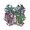
 PDBj
PDBj
 Assembly

 Mass: 851.607 Da / Num. of mol.: 5 / Source method: obtained synthetically / Formula: C25H40N7O18P3S
 Mass: 18.015 Da / Num. of mol.: 595 / Source method: isolated from a natural source / Formula: H2O
 Sample preparation
 / Beamline: X11 / Wavelength: 0.857
  Processing