 Movie
Movie Controller


+ Open data

- Basic information
| Entry | Database: PDB / ID: 1du2 | ||||||
|---|---|---|---|---|---|---|---|
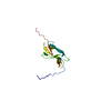
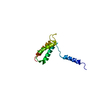
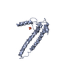
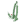
| Title | SOLUTION STRUCTURE OF THE THETA SUBUNIT OF DNA POLYMERASE III | ||||||
 Components Components | DNA POLYMERASE III DNA polymerase III holoenzyme DNA polymerase III holoenzyme | ||||||
 Keywords Keywords | TRANSFERASE / DNA polymerase / Alpha Helix | ||||||
| Function / homology |  Function and homology informationDNA polymerase III, core complex / DNA polymerase III complex / lagging strand elongation / replisome / leading strand elongation / DNA-templated DNA replication / DNA-directed DNA polymerase / DNA-directed DNA polymerase activity / DNA binding / cytosol Function and homology informationDNA polymerase III, core complex / DNA polymerase III complex / lagging strand elongation / replisome / leading strand elongation / DNA-templated DNA replication / DNA-directed DNA polymerase / DNA-directed DNA polymerase activity / DNA binding / cytosolSimilarity search - Function | ||||||
| Biological species |  Escherichia coli (E. coli) Escherichia coli (E. coli) | ||||||
| Method | SOLUTION NMR / torsion angle dynamics | ||||||
 Authors Authors | Keniry, M.A. / Berthon, H.A. / Yang, J.-Y. / Miles, C.S. / Dixon, N.E. | ||||||
 Citation Citation | Journal: Protein Sci. / Year: 2000 Title: NMR solution structure of the theta subunit of DNA polymerase III from Escherichia coli. Authors: Keniry, M.A. / Berthon, H.A. / Yang, J.Y. / Miles, C.S. / Dixon, N.E. | ||||||
| History |
|
- Structure visualization
| Structure viewer | Molecule: MolmilJmol/JSmol |
|---|
- Downloads & links
-Download
| PDBx/mmCIF format | 1du2.cif.gz | 488.7 KB | Display | PDBx/mmCIF format |
|---|---|---|---|---|
| PDB format | pdb1du2.ent.gz | 422.9 KB | Display | PDB format |
| PDBx/mmJSON format | 1du2.json.gz | Tree view | PDBx/mmJSON format | |
| Others |  Other downloads Other downloads |
-Validation report
| Arichive directory | https://data.pdbj.org/pub/pdb/validation_reports/du/1du2ftp://data.pdbj.org/pub/pdb/validation_reports/du/1du2 | HTTPS FTP |
|---|
-Related structure data
| Similar structure data |
|---|


-Links
 PDBj
PDBj
- Assembly
| Deposited unit | 
| |||||||||
|---|---|---|---|---|---|---|---|---|---|---|
| 1 |
| |||||||||
| NMR ensembles |
|
-Components
| #1: Protein | DNA polymerase III holoenzyme Mass: 8861.305 Da / Num. of mol.: 1 / Fragment: THETA SUBUNIT Source method: isolated from a genetically manipulated source Source: (gene. exp.) Escherichia coli (E. coli) / Plasmid: PCM869 / Production host: Escherichia coli (E. coli) / References: UniProt: P0ABS8, DNA-directed DNA polymerase |
|---|
-Experimental details
-Experiment
| Experiment | Method: SOLUTION NMR | ||||||||||||||||||||||||||||
|---|---|---|---|---|---|---|---|---|---|---|---|---|---|---|---|---|---|---|---|---|---|---|---|---|---|---|---|---|---|
| NMR experiment |
| ||||||||||||||||||||||||||||
| NMR details | Text: The structure was determined by double and triple resonance heteronuclear NMR spectroscopy techniques. There is evidence of substantial conformational exchange in sections of the chain that ...Text: The structure was determined by double and triple resonance heteronuclear NMR spectroscopy techniques. There is evidence of substantial conformational exchange in sections of the chain that severely limit the precision of the structure in these regions of the molecule. Specifically, conformational exchange occurs in the regions D19-M33 and L55-S63. |
- Sample preparation
| Details |
| ||||||||||||||||||||
|---|---|---|---|---|---|---|---|---|---|---|---|---|---|---|---|---|---|---|---|---|---|
| Sample conditions |
| ||||||||||||||||||||
| Crystal grow | *PLUS Method: other / Details: NMR |
-NMR measurement
| NMR spectrometer |
|
|---|
- Processing
| NMR software |
| ||||||||||||||||||||
|---|---|---|---|---|---|---|---|---|---|---|---|---|---|---|---|---|---|---|---|---|---|
| Refinement | Method: torsion angle dynamics / Software ordinal: 1 Details: The structures are based on 819 restraints, 511 are NOE-derived distance constraints, 300 dihedral angle restraints and 8 distance restraints from hydrogen bonds | ||||||||||||||||||||
| NMR representative | Selection criteria: fewest violations | ||||||||||||||||||||
| NMR ensemble | Conformer selection criteria: target function / Conformers calculated total number: 400 / Conformers submitted total number: 20 |
