 Movie
Movie Controller


+ Open data

- Basic information
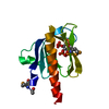
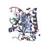
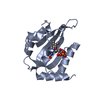
| Entry | Database: PDB / ID: 1dro | ||||||
|---|---|---|---|---|---|---|---|
| Title | NMR STRUCTURE OF THE CYTOSKELETON/SIGNAL TRANSDUCTION PROTEIN | ||||||
 Components Components | BETA-SPECTRIN | ||||||
 Keywords Keywords |  CYTOSKELETON CYTOSKELETON | ||||||
| Function / homology |  Function and homology information Function and homology informationlong-term strengthening of neuromuscular junction / spectrosome / maintenance of presynaptic active zone structure / fusome / photoreceptor cell axon guidance / regulation of plasma membrane organization / COPI-mediated anterograde transport / spectrin / neuromuscular synaptic transmission / negative regulation of microtubule depolymerization ...long-term strengthening of neuromuscular junction / spectrosome / maintenance of presynaptic active zone structure / fusome / photoreceptor cell axon guidance / regulation of plasma membrane organization / COPI-mediated anterograde transport / spectrin / neuromuscular synaptic transmission / negative regulation of microtubule depolymerization / actin filament capping / axon midline choice point recognition / ankyrin binding / regulation of synapse organization / lateral plasma membrane / phosphatidylinositol-4,5-bisphosphate binding / axonogenesis / axon guidance / neuromuscular junction / structural constituent of cytoskeleton / nervous system development / actin binding / microtubule binding / calmodulin binding / axon / plasma membraneSimilarity search - Function | ||||||
| Biological species |  Drosophila melanogaster (fruit fly) Drosophila melanogaster (fruit fly) | ||||||
| Method | SOLUTION NMR | ||||||
 Authors Authors | Zhang, P. / Talluri, S. / Deng, H. / Branton, D. / Wagner, G. | ||||||
 Citation Citation | Journal: Structure / Year: 1995 Title: Solution structure of the pleckstrin homology domain of Drosophila beta-spectrin. Authors: Zhang, P. / Talluri, S. / Deng, H. / Branton, D. / Wagner, G. | ||||||
| History |
|
- Structure visualization
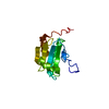
| Structure viewer | Molecule: MolmilJmol/JSmol |
|---|
- Downloads & links
-Download
| PDBx/mmCIF format | 1dro.cif.gz | 558.5 KB | Display | PDBx/mmCIF format |
|---|---|---|---|---|
| PDB format | pdb1dro.ent.gz | 477 KB | Display | PDB format |
| PDBx/mmJSON format | 1dro.json.gz | Tree view | PDBx/mmJSON format | |
| Others |  Other downloads Other downloads |
-Validation report
| Arichive directory | https://data.pdbj.org/pub/pdb/validation_reports/dr/1droftp://data.pdbj.org/pub/pdb/validation_reports/dr/1dro | HTTPS FTP |
|---|
-Related structure data
| Similar structure data |
|---|


-Links
 PDBj
PDBj
- Assembly
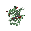
| Deposited unit | 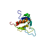
| |||||||||
|---|---|---|---|---|---|---|---|---|---|---|
| 1 |
| |||||||||
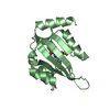
| NMR ensembles |
|
-Components
| #1: Protein | Mass: 13583.059 Da / Num. of mol.: 1 / Fragment: PLECKSTRIN HOMOLOGY DOMAIN Source method: isolated from a genetically manipulated source Source: (gene. exp.) Drosophila melanogaster (fruit fly) / Description: EXPRESSION VECTOR USED WAS PGEX-2T / Organ: FRUIT / Production host:  Escherichia coli (E. coli) / References: UniProt: Q00963 Escherichia coli (E. coli) / References: UniProt: Q00963 |
|---|
-Experimental details
-Experiment
| Experiment | Method: SOLUTION NMR |
|---|
- Sample preparation
| Crystal grow | *PLUS Method: other |
|---|
- Processing
| NMR software | Name: DGII / Developer: HAVEL / Classification: refinement |
|---|---|
| NMR ensemble | Conformers submitted total number: 15 |
