 Movie
Movie Controller


+ Open data

- Basic information
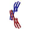
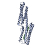
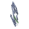
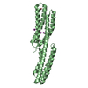
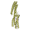
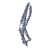
| Entry | Database: PDB / ID: 1dow | ||||||
|---|---|---|---|---|---|---|---|
| Title | CRYSTAL STRUCTURE OF A CHIMERA OF BETA-CATENIN AND ALPHA-CATENIN | ||||||
 Components Components |
| ||||||
 Keywords Keywords |  CELL ADHESION / four-helix bundle CELL ADHESION / four-helix bundle | ||||||
| Function / homology |  Function and homology information Function and homology informationlung cell differentiation / epicardium-derived cardiac vascular smooth muscle cell differentiation / mesenchyme morphogenesis / : / RUNX3 regulates WNT signaling / Regulation of CDH11 function / negative regulation of integrin-mediated signaling pathway / cardiac vascular smooth muscle cell differentiation / Beta-catenin phosphorylation cascade / Disassembly of the destruction complex and recruitment of AXIN to the membrane ...lung cell differentiation / epicardium-derived cardiac vascular smooth muscle cell differentiation / mesenchyme morphogenesis / : / RUNX3 regulates WNT signaling / Regulation of CDH11 function / negative regulation of integrin-mediated signaling pathway / cardiac vascular smooth muscle cell differentiation / Beta-catenin phosphorylation cascade / Disassembly of the destruction complex and recruitment of AXIN to the membrane / Apoptotic cleavage of cell adhesion proteins / hair cycle process / positive regulation of epithelial cell differentiation / TCF dependent signaling in response to WNT / LRR FLII-interacting protein 1 (LRRFIP1) activates type I IFN production / trachea morphogenesis / mesenchyme development / endoderm formation / Formation of the beta-catenin:TCF transactivating complex / VEGFR2 mediated vascular permeability / positive regulation of heparan sulfate proteoglycan biosynthetic process / lung induction / positive regulation of branching involved in lung morphogenesis / cranial ganglion development / renal vesicle formation / renal inner medulla development / renal outer medulla development / nephron tubule formation / mesenchymal stem cell differentiation / beta-catenin-ICAT complex / metanephros morphogenesis / genitalia morphogenesis / embryonic skeletal limb joint morphogenesis / animal organ development / neural plate development / Deactivation of the beta-catenin transactivating complex / glial cell fate determination / regulation of secondary heart field cardioblast proliferation / astrocyte-dopaminergic neuron signaling / negative regulation of mitotic cell cycle, embryonic / canonical Wnt signaling pathway involved in mesenchymal stem cell differentiation / oviduct development / beta-catenin-TCF7L2 complex / regulation of nephron tubule epithelial cell differentiation / negative regulation of mesenchymal to epithelial transition involved in metanephros morphogenesis / regulation of timing of anagen / Ca2+ pathway / central nervous system vasculogenesis / regulation of epithelial cell differentiation / Schwann cell proliferation / regulation of centriole-centriole cohesion / glandular epithelial cell differentiation / Degradation of beta-catenin by the destruction complex / RHO GTPases activate IQGAPs / regulation of centromeric sister chromatid cohesion / Adherens junctions interactions / embryonic axis specification / endodermal cell fate commitment / ventricular compact myocardium morphogenesis / morphogenesis of embryonic epithelium / Scrib-APC-beta-catenin complex / positive regulation of fibroblast growth factor receptor signaling pathway / beta-catenin-TCF complex / lens morphogenesis in camera-type eye / gap junction assembly / dorsal root ganglion development / synaptic vesicle clustering / acinar cell differentiation / epithelial cell-cell adhesion / zonula adherens / dorsal/ventral axis specification / proximal/distal pattern formation / neuron fate determination / layer formation in cerebral cortex / positive regulation of myoblast proliferation / positive regulation of endothelial cell differentiation / sympathetic ganglion development / establishment of blood-retinal barrier / fungiform papilla formation / gamma-catenin binding / lung epithelial cell differentiation / delta-catenin binding / embryonic foregut morphogenesis / hindbrain development / regulation of calcium ion import / positive regulation of determination of dorsal identity / positive regulation of skeletal muscle tissue development / ectoderm development / positive regulation of odontoblast differentiation / regulation of osteoclast differentiation / cranial skeletal system development / endothelial tube morphogenesis / regulation of protein localization to cell surface / mesenchymal cell proliferation involved in lung development / smooth muscle cell differentiation / histone methyltransferase binding / cell projection membrane / midbrain dopaminergic neuron differentiation / presynaptic active zone cytoplasmic component / mesenchymal cell proliferationSimilarity search - Function | ||||||
| Biological species |  Mus musculus (house mouse) Mus musculus (house mouse) | ||||||
| Method | X-RAY DIFFRACTION / SYNCHROTRON / Resolution: 1.8 Å | ||||||
 Authors Authors | Pokutta, S. / Weis, W.I. | ||||||
 Citation Citation | Journal: Mol.Cell / Year: 2000 Title: Structure of the dimerization and beta-catenin-binding region of alpha-catenin. Authors: Pokutta, S. / Weis, W.I. | ||||||
| History |
|
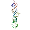
- Structure visualization
| Structure viewer | Molecule: MolmilJmol/JSmol |
|---|
- Downloads & links
-Download
| PDBx/mmCIF format | 1dow.cif.gz | 61.4 KB | Display | PDBx/mmCIF format |
|---|---|---|---|---|
| PDB format | pdb1dow.ent.gz | 47 KB | Display | PDB format |
| PDBx/mmJSON format | 1dow.json.gz | Tree view | PDBx/mmJSON format | |
| Others |  Other downloads Other downloads |
-Validation report
| Arichive directory | https://data.pdbj.org/pub/pdb/validation_reports/do/1dowftp://data.pdbj.org/pub/pdb/validation_reports/do/1dow | HTTPS FTP |
|---|
-Related structure data


-Links
 PDBj
PDBj


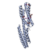
- Assembly
| Deposited unit | 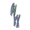
| ||||||||
|---|---|---|---|---|---|---|---|---|---|
| 1 |
| ||||||||
| Unit cell |
|
-Components
| #1: Protein | Alpha catenin Mass: 22694.391 Da / Num. of mol.: 1 / Fragment: DIMERIZATION AND BETA-CATENIN BINDING REGION Source method: isolated from a genetically manipulated source Source: (gene. exp.) Mus musculus (house mouse) / Production host:  Escherichia coli (E. coli) / References: UniProt: P26231 Escherichia coli (E. coli) / References: UniProt: P26231 |
|---|---|
| #2: Protein/peptide | Catenin beta-1 Mass: 3666.949 Da / Num. of mol.: 1 / Fragment: ALPHA-CATENIN BINDING REGION Source method: isolated from a genetically manipulated source Source: (gene. exp.) Mus musculus (house mouse) / Production host: Escherichia coli (E. coli) / References: UniProt: Q02248 |
| #3: Water | ChemComp-HOH / Water Mass: 18.015 Da / Num. of mol.: 252 / Source method: isolated from a natural source / Formula: H2O Mass: 18.015 Da / Num. of mol.: 252 / Source method: isolated from a natural source / Formula: H2O |
-Experimental details
-Experiment
| Experiment | Method: X-RAY DIFFRACTION / Number of used crystals: 1 |
|---|
- Sample preparation
| Crystal | Density Matthews: 2 Å3/Da / Density % sol: 38.6 % | |||||||||||||||||||||||||||||||||||
|---|---|---|---|---|---|---|---|---|---|---|---|---|---|---|---|---|---|---|---|---|---|---|---|---|---|---|---|---|---|---|---|---|---|---|---|---|
| Crystal grow | Temperature: 295 K / Method: vapor diffusion, hanging drop / pH: 7 Details: PEG 2000 monomethylether, HEPES, urea, ethanol, Dithiothreitol, pH 7.0, VAPOR DIFFUSION, HANGING DROP, temperature 295.0K | |||||||||||||||||||||||||||||||||||
| Crystal grow | *PLUS | |||||||||||||||||||||||||||||||||||
| Components of the solutions | *PLUS
|
-Data collection
| Diffraction | Mean temperature: 100 K |
|---|---|
| Diffraction source | Source: SYNCHROTRON / Site: SSRL  / Beamline: BL1-5 / Wavelength: 0.9252 / Beamline: BL1-5 / Wavelength: 0.9252 |
| Detector | Type: ADSC QUANTUM 4 / Detector: CCD / Date: Jan 1, 1999 |
| Radiation | Protocol: SINGLE WAVELENGTH / Monochromatic (M) / Laue (L): M / Scattering type: x-ray |
| Radiation wavelength | Wavelength: 0.9252 Å / Relative weight: 1 |
| Reflection | Resolution: 1.8→30 Å / Num. all: 37629 / Num. obs: 36760 / % possible obs: 99.3 % / Observed criterion σ(F): 0 / Observed criterion σ(I): -3 / Redundancy: 3.2 % / Biso Wilson estimate: 21.5 Å2 / Rmerge(I) obs: 0.042 / Net I/σ(I): 28.4 |
| Reflection shell | Resolution: 1.8→1.83 Å / Redundancy: 2.5 % / Rmerge(I) obs: 0.212 / Num. unique all: 1759 / % possible all: 92.3 |
| Reflection shell | *PLUS % possible obs: 92.3 % |
- Processing
| Software |
| ||||||||||||||||||||||||||||||||||||
|---|---|---|---|---|---|---|---|---|---|---|---|---|---|---|---|---|---|---|---|---|---|---|---|---|---|---|---|---|---|---|---|---|---|---|---|---|---|
| Refinement | Resolution: 1.8→30 Å / Rfactor Rfree error: 0.004 / Data cutoff high absF: 309534.83 / Data cutoff low absF: 0 / Isotropic thermal model: RESTRAINED / Cross valid method: THROUGHOUT / σ(F): 0 / Stereochemistry target values: ENGH AND HUBER
| ||||||||||||||||||||||||||||||||||||
| Solvent computation | Solvent model: FLAT MODEL / Bsol: 38.93 Å2 / ksol: 0.336 e/Å3 | ||||||||||||||||||||||||||||||||||||
| Displacement parameters | Biso mean: 26.1 Å2
| ||||||||||||||||||||||||||||||||||||
| Refine analyze |
| ||||||||||||||||||||||||||||||||||||
| Refinement step | Cycle: LAST / Resolution: 1.8→30 Å
| ||||||||||||||||||||||||||||||||||||
| Refine LS restraints |
| ||||||||||||||||||||||||||||||||||||
| LS refinement shell | Resolution: 1.8→1.86 Å / Rfactor Rfree error: 0.016 / Total num. of bins used: 10
| ||||||||||||||||||||||||||||||||||||
| Xplor file |
| ||||||||||||||||||||||||||||||||||||
| Software | *PLUS Name: CNS / Version: 0.5 / Classification: refinement | ||||||||||||||||||||||||||||||||||||
| Refinement | *PLUS Num. reflection all: 33897 | ||||||||||||||||||||||||||||||||||||
| Solvent computation | *PLUS | ||||||||||||||||||||||||||||||||||||
| Displacement parameters | *PLUS | ||||||||||||||||||||||||||||||||||||
| Refine LS restraints | *PLUS
|
