 Movie
Movie Controller

[English] 日本語
 Yorodumi
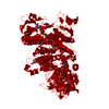
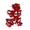
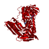
- PDB-1dnc: HUMAN GLUTATHIONE REDUCTASE MODIFIED BY DIGLUTATHIONE-DINITROSO-IRON -
+ Open data

- Basic information
| Entry | Database: PDB / ID: 1dnc | ||||||
|---|---|---|---|---|---|---|---|
| Title | HUMAN GLUTATHIONE REDUCTASE MODIFIED BY DIGLUTATHIONE-DINITROSO-IRON | ||||||
 Components Components | GLUTATHIONE REDUCTASE | ||||||
 Keywords Keywords | OXIDOREDUCTASE / SULFHYDRYL OXIDATION / SULFINIC ACID / NITRIC OXIDE | ||||||
| Function / homology |  Function and homology informationglutathione-disulfide reductase / Metabolism of ingested H2SeO4 and H2SeO3 into H2Se / glutathione-disulfide reductase (NADPH) activity / Interconversion of nucleotide di- and triphosphates / NFE2L2 regulating anti-oxidant/detoxification enzymes / Detoxification of Reactive Oxygen Species / glutathione metabolic process / cell redox homeostasis / TP53 Regulates Metabolic Genes / flavin adenine dinucleotide binding ...glutathione-disulfide reductase / Metabolism of ingested H2SeO4 and H2SeO3 into H2Se / glutathione-disulfide reductase (NADPH) activity / Interconversion of nucleotide di- and triphosphates / NFE2L2 regulating anti-oxidant/detoxification enzymes / Detoxification of Reactive Oxygen Species / glutathione metabolic process / cell redox homeostasis / TP53 Regulates Metabolic Genes / flavin adenine dinucleotide binding / cellular response to oxidative stress / NADP binding / electron transfer activity / mitochondrial matrix / external side of plasma membrane / mitochondrion / extracellular exosome / cytosol Function and homology informationglutathione-disulfide reductase / Metabolism of ingested H2SeO4 and H2SeO3 into H2Se / glutathione-disulfide reductase (NADPH) activity / Interconversion of nucleotide di- and triphosphates / NFE2L2 regulating anti-oxidant/detoxification enzymes / Detoxification of Reactive Oxygen Species / glutathione metabolic process / cell redox homeostasis / TP53 Regulates Metabolic Genes / flavin adenine dinucleotide binding ...glutathione-disulfide reductase / Metabolism of ingested H2SeO4 and H2SeO3 into H2Se / glutathione-disulfide reductase (NADPH) activity / Interconversion of nucleotide di- and triphosphates / NFE2L2 regulating anti-oxidant/detoxification enzymes / Detoxification of Reactive Oxygen Species / glutathione metabolic process / cell redox homeostasis / TP53 Regulates Metabolic Genes / flavin adenine dinucleotide binding / cellular response to oxidative stress / NADP binding / electron transfer activity / mitochondrial matrix / external side of plasma membrane / mitochondrion / extracellular exosome / cytosolSimilarity search - Function | ||||||
| Biological species |  Homo sapiens (human) Homo sapiens (human) | ||||||
| Method | X-RAY DIFFRACTION / Resolution: 1.7 Å | ||||||
 Authors Authors | Becker, K. / Savvides, S.N. / Keese, M. / Schirmer, R.H. / Karplus, P.A. | ||||||
 Citation Citation | Journal: Nat.Struct.Biol. / Year: 1998 Title: Enzyme inactivation through sulfhydryl oxidation by physiologic NO-carriers. Authors: Becker, K. / Savvides, S.N. / Keese, M. / Schirmer, R.H. / Karplus, P.A. | ||||||
| History |
|
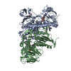
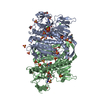
- Structure visualization
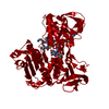
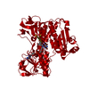
| Structure viewer | Molecule: MolmilJmol/JSmol |
|---|
- Downloads & links
-Download
| PDBx/mmCIF format | 1dnc.cif.gz | 111.6 KB | Display | PDBx/mmCIF format |
|---|---|---|---|---|
| PDB format | pdb1dnc.ent.gz | 90.1 KB | Display | PDB format |
| PDBx/mmJSON format | 1dnc.json.gz | Tree view | PDBx/mmJSON format | |
| Others |  Other downloads Other downloads |
-Validation report
| Arichive directory | https://data.pdbj.org/pub/pdb/validation_reports/dn/1dncftp://data.pdbj.org/pub/pdb/validation_reports/dn/1dnc | HTTPS FTP |
|---|
-Related structure data


-Links
 PDBj
PDBj


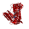
- Assembly
| Deposited unit | 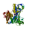
| ||||||||
|---|---|---|---|---|---|---|---|---|---|
| 1 | 
| ||||||||
| Unit cell |
|
-Components
| #1: Protein | Mass: 51668.242 Da / Num. of mol.: 1 Source method: isolated from a genetically manipulated source Details: MIXED DISULFIDE BETWEEN C58 AND GLUTATHIONE (GSH 1029) SULFINIC ACID GROUP IN CIA63 Source: (gene. exp.) Homo sapiens (human) / Production host:  Escherichia coli (E. coli) / References: UniProt: P00390, EC: 1.6.4.2 Escherichia coli (E. coli) / References: UniProt: P00390, EC: 1.6.4.2 |
|---|---|
| #2: Chemical | ChemComp-PO4 / Phosphate  Mass: 94.971 Da / Num. of mol.: 1 / Source method: obtained synthetically / Formula: PO4 Mass: 94.971 Da / Num. of mol.: 1 / Source method: obtained synthetically / Formula: PO4 |
| #3: Chemical | ChemComp-FAD / Flavin adenine dinucleotide  Mass: 785.550 Da / Num. of mol.: 1 / Source method: obtained synthetically / Formula: C27H33N9O15P2 / Comment: FAD*YM Mass: 785.550 Da / Num. of mol.: 1 / Source method: obtained synthetically / Formula: C27H33N9O15P2 / Comment: FAD*YM |
| #4: Chemical | ChemComp-GSH / Glutathione  Mass: 307.323 Da / Num. of mol.: 1 / Source method: obtained synthetically / Formula: C10H17N3O6S Mass: 307.323 Da / Num. of mol.: 1 / Source method: obtained synthetically / Formula: C10H17N3O6S |
| #5: Water | ChemComp-HOH / Water Mass: 18.015 Da / Num. of mol.: 518 / Source method: isolated from a natural source / Formula: H2O Mass: 18.015 Da / Num. of mol.: 518 / Source method: isolated from a natural source / Formula: H2O |
-Experimental details
-Experiment
| Experiment | Method: X-RAY DIFFRACTION |
|---|
- Sample preparation
| Crystal | Density Matthews: 2.65 Å3/Da / Density % sol: 53.66 % | |||||||||||||||||||||||||||||||||||
|---|---|---|---|---|---|---|---|---|---|---|---|---|---|---|---|---|---|---|---|---|---|---|---|---|---|---|---|---|---|---|---|---|---|---|---|---|
| Crystal grow | *PLUS pH: 7.5 / Method: vapor diffusion, hanging drop | |||||||||||||||||||||||||||||||||||
| Components of the solutions | *PLUS
|
-Data collection
| Diffraction | Mean temperature: 298 K |
|---|---|
| Diffraction source | Wavelength: 1.5418 |
| Detector | Type: XUONG-HAMLIN MULTIWIRE / Detector: AREA DETECTOR / Date: Aug 1, 1996 |
| Radiation | Monochromatic (M) / Laue (L): M / Scattering type: x-ray |
| Radiation wavelength | Wavelength: 1.5418 Å / Relative weight: 1 |
| Reflection | Highest resolution: 1.7 Å / Num. obs: 58100 / % possible obs: 97 % / Redundancy: 4.1 % / Rsym value: 0.093 / Net I/σ(I): 14.7 |
| Reflection shell | Resolution: 1.7→1.74 Å / Mean I/σ(I) obs: 2 / Rsym value: 0.329 / % possible all: 75 |
| Reflection | *PLUS Num. measured all: 239537 / Rmerge(I) obs: 0.093 |
| Reflection shell | *PLUS % possible obs: 75 % / Rmerge(I) obs: 0.329 |
- Processing
| Software |
| ||||||||||||||||||||||||||||||||||||||||||||||||||||||||||||
|---|---|---|---|---|---|---|---|---|---|---|---|---|---|---|---|---|---|---|---|---|---|---|---|---|---|---|---|---|---|---|---|---|---|---|---|---|---|---|---|---|---|---|---|---|---|---|---|---|---|---|---|---|---|---|---|---|---|---|---|---|---|
| Refinement | Resolution: 1.7→10 Å / Cross valid method: THROUGHOUT / σ(F): 1
| ||||||||||||||||||||||||||||||||||||||||||||||||||||||||||||
| Refinement step | Cycle: LAST / Resolution: 1.7→10 Å
| ||||||||||||||||||||||||||||||||||||||||||||||||||||||||||||
| Refine LS restraints |
| ||||||||||||||||||||||||||||||||||||||||||||||||||||||||||||
| Software | *PLUS Name: X-PLOR / Version: 3.1 / Classification: refinement | ||||||||||||||||||||||||||||||||||||||||||||||||||||||||||||
| Refinement | *PLUS | ||||||||||||||||||||||||||||||||||||||||||||||||||||||||||||
| Solvent computation | *PLUS | ||||||||||||||||||||||||||||||||||||||||||||||||||||||||||||
| Displacement parameters | *PLUS |
