 Movie
Movie Controller


+ Open data

- Basic information
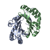
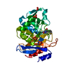
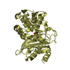
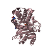
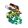
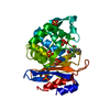
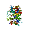
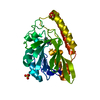
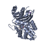
| Entry | Database: PDB / ID: 1dja | ||||||
|---|---|---|---|---|---|---|---|
| Title | STRUCTURE OF BETA-LACTAMASE PRECURSOR, K73H MUTANT, AT 298K | ||||||
 Components Components | BETA-LACTAMASE | ||||||
 Keywords Keywords | HYDROLASE / ANTIBIOTIC RESISTANCE / PLASMID / TRANSPOSABLE ELEMENT | ||||||
| Function / homology |  Function and homology information Function and homology informationbeta-lactam antibiotic catabolic process / beta-lactamase activity / beta-lactamase / response to antibioticSimilarity search - Function | ||||||
| Biological species |   Staphylococcus aureus (bacteria) Staphylococcus aureus (bacteria) | ||||||
| Method | X-RAY DIFFRACTION / Resolution: 1.9 Å | ||||||
 Authors Authors | Chen, C.C.H. / Herzberg, O. | ||||||
 Citation Citation | Journal: Biochemistry / Year: 1996 Title: Structure and kinetics of the beta-lactamase mutants S70A and K73H from Staphylococcus aureus PC1. Authors: Chen, C.C. / Smith, T.J. / Kapadia, G. / Wasch, S. / Zawadzke, L.E. / Coulson, A. / Herzberg, O. #1: Journal: Protein Eng. / Year: 1995Title: An Engineered Staphylococcus Aureus Pc1 Beta-Lactamase that Hydrolyses Third-Generation Cephalosporins Authors: Zawadzke, L.E. / Smith, T.J. / Herzberg, O. #2: Journal: J.Mol.Biol. / Year: 1991Title: Refined Crystal Structure of Beta-Lactamase from Staphylococcus Aureus Pc1 at 2.0 Authors: Herzberg, O. #3: Journal: Science / Year: 1987Title: Bacterial Resistance to Beta-Lactam Antibiotics. Crystal Structure of Beta-Lactamase from Staphylococcus Aureus Pc1 at 2.5 A Resolution Authors: Herzberg, O. / Moult, J. | ||||||
| History |
|
- Structure visualization
| Structure viewer | Molecule: MolmilJmol/JSmol |
|---|
- Downloads & links
-Download
| PDBx/mmCIF format | 1dja.cif.gz | 65.9 KB | Display | PDBx/mmCIF format |
|---|---|---|---|---|
| PDB format | pdb1dja.ent.gz | 47.9 KB | Display | PDB format |
| PDBx/mmJSON format | 1dja.json.gz | Tree view | PDBx/mmJSON format | |
| Others |  Other downloads Other downloads |
-Validation report
| Arichive directory | https://data.pdbj.org/pub/pdb/validation_reports/dj/1djaftp://data.pdbj.org/pub/pdb/validation_reports/dj/1dja | HTTPS FTP |
|---|
-Related structure data


-Links
 PDBj
PDBj


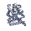
- Assembly
| Deposited unit | 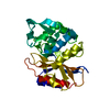
| |||||||||
|---|---|---|---|---|---|---|---|---|---|---|
| 1 |
| |||||||||
| Unit cell |
| |||||||||
| Components on special symmetry positions |
|
-Components
| #1: Protein | / PENICILLINASE Mass: 28983.375 Da / Num. of mol.: 1 / Mutation: A30M, K73H Source method: isolated from a genetically manipulated source Source: (gene. exp.) Staphylococcus aureus (bacteria) / Strain: PC1 / Description: INDUCIBLE TRC PROMOTER / Gene: BLAZ / Plasmid: PTS32 / Gene (production host): BLAZ / Production host: Escherichia coli (E. coli) / References: UniProt: P00807, beta-lactamase |
|---|---|
| #2: Water | ChemComp-HOH / Water Mass: 18.015 Da / Num. of mol.: 177 / Source method: isolated from a natural source / Formula: H2O Mass: 18.015 Da / Num. of mol.: 177 / Source method: isolated from a natural source / Formula: H2O |
-Experimental details
-Experiment
| Experiment | Method: X-RAY DIFFRACTION |
|---|
- Sample preparation
| Crystal | Density Matthews: 3.03 Å3/Da / Density % sol: 65 % | ||||||||||||||||||||||||||||||||||||
|---|---|---|---|---|---|---|---|---|---|---|---|---|---|---|---|---|---|---|---|---|---|---|---|---|---|---|---|---|---|---|---|---|---|---|---|---|---|
| Crystal grow | *PLUS pH: 8 / Method: vapor diffusion, hanging dropDetails: drop consists of equal volume of protein and reservoir solutions | ||||||||||||||||||||||||||||||||||||
| Components of the solutions | *PLUS
|
-Data collection
| Diffraction | Mean temperature: 298 K |
|---|---|
| Diffraction source | Wavelength: 1.5418 |
| Detector | Type: SIEMENS / Detector: AREA DETECTOR / Date: Nov 6, 1995 |
| Radiation | Monochromatic (M) / Laue (L): M / Scattering type: x-ray |
| Radiation wavelength | Wavelength: 1.5418 Å / Relative weight: 1 |
| Reflection | Num. obs: 21589 / % possible obs: 79 % / Observed criterion σ(I): 0 / Redundancy: 2.9 % / Rmerge(I) obs: 0.067 |
| Reflection | *PLUS Highest resolution: 1.9 Å / Num. measured all: 61878 |
| Reflection shell | *PLUS % possible obs: 44 % / Num. unique obs: 1971 / Num. measured obs: 3897 / Rmerge(I) obs: 0.218 |
- Processing
| Software |
| ||||||||||||||||||||||||||||||||||||||||||||||||||||||||||||
|---|---|---|---|---|---|---|---|---|---|---|---|---|---|---|---|---|---|---|---|---|---|---|---|---|---|---|---|---|---|---|---|---|---|---|---|---|---|---|---|---|---|---|---|---|---|---|---|---|---|---|---|---|---|---|---|---|---|---|---|---|---|
| Refinement | Resolution: 1.9→8 Å / σ(F): 2 /
| ||||||||||||||||||||||||||||||||||||||||||||||||||||||||||||
| Refinement step | Cycle: LAST / Resolution: 1.9→8 Å
| ||||||||||||||||||||||||||||||||||||||||||||||||||||||||||||
| Refine LS restraints |
|
