[English] 日本語
 Yorodumi
- PDB-1deq: THE CRYSTAL STRUCTURE OF MODIFIED BOVINE FIBRINOGEN (AT ~4 ANGSTR... -
+ Open data
- Basic information
| Entry | Database: PDB / ID: 1deq | ||||||
|---|---|---|---|---|---|---|---|
| Title | THE CRYSTAL STRUCTURE OF MODIFIED BOVINE FIBRINOGEN (AT ~4 ANGSTROM RESOLUTION) | ||||||
 Components Components |
| ||||||
 Keywords Keywords |  BLOOD CLOTTING / BLOOD CLOTTING /  COILED-COIL COILED-COIL | ||||||
| Function / homology |  Function and homology information Function and homology information blood coagulation, common pathway / blood coagulation, common pathway /  fibrinogen complex / fibrinogen complex /  blood coagulation, fibrin clot formation / positive regulation of heterotypic cell-cell adhesion / positive regulation of peptide hormone secretion / protein polymerization / negative regulation of endothelial cell apoptotic process / negative regulation of extrinsic apoptotic signaling pathway via death domain receptors / blood coagulation, fibrin clot formation / positive regulation of heterotypic cell-cell adhesion / positive regulation of peptide hormone secretion / protein polymerization / negative regulation of endothelial cell apoptotic process / negative regulation of extrinsic apoptotic signaling pathway via death domain receptors /  fibrinolysis / cell-matrix adhesion ... fibrinolysis / cell-matrix adhesion ... blood coagulation, common pathway / blood coagulation, common pathway /  fibrinogen complex / fibrinogen complex /  blood coagulation, fibrin clot formation / positive regulation of heterotypic cell-cell adhesion / positive regulation of peptide hormone secretion / protein polymerization / negative regulation of endothelial cell apoptotic process / negative regulation of extrinsic apoptotic signaling pathway via death domain receptors / blood coagulation, fibrin clot formation / positive regulation of heterotypic cell-cell adhesion / positive regulation of peptide hormone secretion / protein polymerization / negative regulation of endothelial cell apoptotic process / negative regulation of extrinsic apoptotic signaling pathway via death domain receptors /  fibrinolysis / cell-matrix adhesion / fibrinolysis / cell-matrix adhesion /  platelet aggregation / collagen-containing extracellular matrix / platelet aggregation / collagen-containing extracellular matrix /  adaptive immune response / positive regulation of ERK1 and ERK2 cascade / adaptive immune response / positive regulation of ERK1 and ERK2 cascade /  signaling receptor binding / signaling receptor binding /  innate immune response / innate immune response /  extracellular space / extracellular space /  metal ion binding metal ion bindingSimilarity search - Function | ||||||
| Biological species |   Bos taurus (cattle) Bos taurus (cattle) | ||||||
| Method |  X-RAY DIFFRACTION / X-RAY DIFFRACTION /  SYNCHROTRON / molecular replacement (using human fragment d coordinates) / Resolution: 3.5 Å SYNCHROTRON / molecular replacement (using human fragment d coordinates) / Resolution: 3.5 Å | ||||||
 Authors Authors | Brown, J.H. / Volkmann, N. / Jun, G. / Henschen-Edman, A.H. / Cohen, C. | ||||||
 Citation Citation |  Journal: Proc.Natl.Acad.Sci.USA / Year: 2000 Journal: Proc.Natl.Acad.Sci.USA / Year: 2000Title: The crystal structure of modified bovine fibrinogen. Authors: Brown, J.H. / Volkmann, N. / Jun, G. / Henschen-Edman, A.H. / Cohen, C. #1:  Journal: J.Mol.Biol. / Year: 1991 Journal: J.Mol.Biol. / Year: 1991Title: Fibrinogen Structure in Projection at 18 Angstroms Resolution Authors: Rao, S.P.S. / Poojary, M.D. / Elliott Jr., B.W. / Melanson, L.A. / Oriel, B. / Cohen, C. #2:  Journal: J.Mol.Biol. / Year: 1978 Journal: J.Mol.Biol. / Year: 1978Title: Crystals of Modified Fibrinogen: Size, Shape and Packing of Molecules Authors: Weisel, J.W. / Warren, S.G. / Cohen, C. #3:  Journal: J.Mol.Biol. / Year: 1977 Journal: J.Mol.Biol. / Year: 1977Title: Crystalline States of a Modified Fibrinogen Authors: Tooney, N.M. / Cohen, C. | ||||||
| History |
|
- Structure visualization
| Structure viewer | Molecule:  Molmil Molmil Jmol/JSmol Jmol/JSmol |
|---|
- Downloads & links
- Download
| PDBx/mmCIF format |  1deq.cif.gz 1deq.cif.gz | 161.2 KB | Display |  PDBx/mmCIF format PDBx/mmCIF format |
|---|---|---|---|---|
| PDB format |  pdb1deq.ent.gz pdb1deq.ent.gz | 89.3 KB | Display |  PDB format PDB format |
| PDBx/mmJSON format |  1deq.json.gz 1deq.json.gz | Tree view |  PDBx/mmJSON format PDBx/mmJSON format | |
| Others |  Other downloads Other downloads |
-Validation report
| Arichive directory |  https://data.pdbj.org/pub/pdb/validation_reports/de/1deq https://data.pdbj.org/pub/pdb/validation_reports/de/1deq ftp://data.pdbj.org/pub/pdb/validation_reports/de/1deq ftp://data.pdbj.org/pub/pdb/validation_reports/de/1deq | HTTPS FTP |
|---|
-Related structure data
| Related structure data |  1fzaS S: Starting model for refinement |
|---|---|
| Similar structure data |
- Links
Links
- Assembly
| Deposited unit | 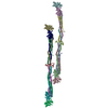
| ||||||||
|---|---|---|---|---|---|---|---|---|---|
| 1 | 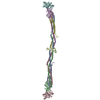
| ||||||||
| 2 | 
| ||||||||
| Unit cell |
| ||||||||
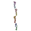
| Details | An ~ 4 Angstrom structure of the 285 kDa major fragment of bovine fibrinogen. The alpha-carbon coordinates for both molecules per asymmetric unit are provided. The molecule is a dimer of a heterotrimer. Coordinates with chain ids ABC:DEF and NOP:QRS are the two crystallographically independent dimers. A,D,N, and Q correspond to the A-alpha chain. B,E,O, and R correspond to the B-beta chain. C,F,P, and S correspond to the gamma chain. These alpha-carbon coordinates include the end gamma and beta domains (i.e. the C-terminal globular domains of the gamma and B-beta chains, respectively) and the coiled coils (which are made of the A-alpha, B-beta, and gamma chains.). The density corresponding to the central disulfide knot region is too irregular to be traced at this resolution, and only some coordinates for part of this domain are included where density is seen (designated M and Z for the for the two molecules per a.u.). |
- Components
| #1: Protein | Mass: 42767.410 Da / Num. of mol.: 4 / Fragment: PSEUDOMONAS AERUGINOSA PS-1-MODIFIED FRAGMENT / Source method: isolated from a natural source / Source: (natural)   Bos taurus (cattle) / References: UniProt: P02672 Bos taurus (cattle) / References: UniProt: P02672#2: Protein | Mass: 46901.641 Da / Num. of mol.: 4 / Fragment: PSEUDOMONAS AERUGINOSA PS-1-MODIFIED FRAGMENT / Source method: isolated from a natural source / Source: (natural)   Bos taurus (cattle) / References: UniProt: P02676 Bos taurus (cattle) / References: UniProt: P02676#3: Protein | Mass: 46626.605 Da / Num. of mol.: 4 / Fragment: PSEUDOMONAS AERUGINOSA PS-1-MODIFIED FRAGMENT / Source method: isolated from a natural source / Source: (natural)   Bos taurus (cattle) / References: UniProt: P12799 Bos taurus (cattle) / References: UniProt: P12799#4: Protein |  / Coordinate model: Cα atoms only / Coordinate model: Cα atoms onlyMass: 7677.455 Da / Num. of mol.: 2 / Fragment: PSEUDOMONAS AERUGINOSA PS-1-MODIFIED FRAGMENT / Source method: isolated from a natural source / Details: DISORDERED DISULFIDE KNOT REGION / Source: (natural)   Bos taurus (cattle) Bos taurus (cattle) |
|---|
-Experimental details
-Experiment
| Experiment | Method:  X-RAY DIFFRACTION / Number of used crystals: 3 X-RAY DIFFRACTION / Number of used crystals: 3 |
|---|
- Sample preparation
| Crystal | Density Matthews: 3.12 Å3/Da / Density % sol: 60.53 % | ||||||||||||||||||||||||||||||
|---|---|---|---|---|---|---|---|---|---|---|---|---|---|---|---|---|---|---|---|---|---|---|---|---|---|---|---|---|---|---|---|
Crystal grow | Temperature: 277 K / pH: 6.2 Details: 10 mM MES, 5mM sodium azide, 2mM calcium chloride , pH 6.2, temperature 277K | ||||||||||||||||||||||||||||||
| Crystal grow | *PLUS Method: batch method | ||||||||||||||||||||||||||||||
| Components of the solutions | *PLUS
|
-Data collection
| Diffraction |
| ||||||||||||||||||||
|---|---|---|---|---|---|---|---|---|---|---|---|---|---|---|---|---|---|---|---|---|---|
| Diffraction source |
| ||||||||||||||||||||
| Detector |
| ||||||||||||||||||||
| Radiation | Protocol: SINGLE WAVELENGTH / Monochromatic (M) / Laue (L): M / Scattering type: x-ray | ||||||||||||||||||||
| Radiation wavelength |
| ||||||||||||||||||||
| Reflection | Resolution: 3.4→210 Å / Num. all: 79465 / Num. obs: 79465 / % possible obs: 78.5 % / Observed criterion σ(F): 0 / Observed criterion σ(I): 0 / Redundancy: 4.42 % / Rmerge(I) obs: 0.075 / Net I/σ(I): 7.05 | ||||||||||||||||||||
| Reflection shell | Resolution: 3.38→3.57 Å / Redundancy: 2.3 % / Rmerge(I) obs: 0.28 / Num. unique all: 6830 / % possible all: 46.7 | ||||||||||||||||||||
| Reflection | *PLUS | ||||||||||||||||||||
| Reflection shell | *PLUS % possible obs: 46.7 % |
- Processing
| Software |
| |||||||||||||||||||||||||
|---|---|---|---|---|---|---|---|---|---|---|---|---|---|---|---|---|---|---|---|---|---|---|---|---|---|---|
| Refinement | Method to determine structure : molecular replacement (using human fragment d coordinates) : molecular replacement (using human fragment d coordinates)Starting model: human fragment d coordinates (see related entry 1fza) Resolution: 3.5→10 Å / σ(F): 0 / Stereochemistry target values: modified CHARMM in X-Plor Details: Side chains were included in refinement. At this resolution, however, the correct side chain conformations cannot be generally determined, and only alpha Carbons are deposited here. A B- ...Details: Side chains were included in refinement. At this resolution, however, the correct side chain conformations cannot be generally determined, and only alpha Carbons are deposited here. A B-factor equal to 999 identifies a residue whose density is generally relatively disordered and thus is not included in refinement (but generally positioned using information from non- crystallographically-related copies). Note that of the asymmetric unit's four half-dimers, that designated by chain ids N, O, and P has the most residues included in the refinement, and its electron density is best ordered. The central disulphide knot region could not be traced, and only some coordinates for part of this domain are included (resname=UNK, chainid M and Z for the two molecules per a.u.). Due to anisotropy in the the diffraction, the data in the highest shells (esp. 4.0-3.5) are quite incomplete, and hence the structure is judged overall to at ~4.0 angstroms resolution.
| |||||||||||||||||||||||||
| Refinement step | Cycle: LAST / Resolution: 3.5→10 Å
| |||||||||||||||||||||||||
| Refine LS restraints |
| |||||||||||||||||||||||||
| Software | *PLUS Name:  X-PLOR / Version: 3.851 / Classification: refinement X-PLOR / Version: 3.851 / Classification: refinement | |||||||||||||||||||||||||
| Refine LS restraints | *PLUS
|
 Movie
Movie Controller


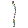

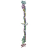
 PDBj
PDBj


