 Movie
Movie Controller


+ Open data

- Basic information
| Entry | Database: PDB / ID: 1d5s | ||||||
|---|---|---|---|---|---|---|---|
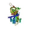
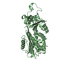
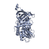
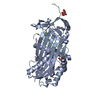
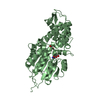
| Title | CRYSTAL STRUCTURE OF CLEAVED ANTITRYPSIN POLYMER | ||||||
 Components Components | (P1-ARG ANTITRYPSIN) x 2 | ||||||
 Keywords Keywords | HYDROLASE INHIBITOR / SERPIN FOLD / RCL CLEAVAGE / A BETA SHEET POLYMERISATION | ||||||
| Function / homology |  Function and homology information Function and homology informationCargo concentration in the ER / COPII-mediated vesicle transport / COPII-coated ER to Golgi transport vesicle / endoplasmic reticulum-Golgi intermediate compartment membrane / platelet alpha granule lumen / acute-phase response /  Post-translational protein phosphorylation / serine-type endopeptidase inhibitor activity / Regulation of Insulin-like Growth Factor (IGF) transport and uptake by Insulin-like Growth Factor Binding Proteins (IGFBPs) / blood coagulation ...Cargo concentration in the ER / COPII-mediated vesicle transport / COPII-coated ER to Golgi transport vesicle / endoplasmic reticulum-Golgi intermediate compartment membrane / platelet alpha granule lumen / acute-phase response / Post-translational protein phosphorylation / serine-type endopeptidase inhibitor activity / Regulation of Insulin-like Growth Factor (IGF) transport and uptake by Insulin-like Growth Factor Binding Proteins (IGFBPs) / blood coagulation / Platelet degranulation / collagen-containing extracellular matrix / protease binding / ficolin-1-rich granule lumen / endoplasmic reticulum lumen / intracellular membrane-bounded organelle / Neutrophil degranulation / Golgi apparatus / endoplasmic reticulum / extracellular space / extracellular exosome / extracellular region / identical protein binding Post-translational protein phosphorylation / serine-type endopeptidase inhibitor activity / Regulation of Insulin-like Growth Factor (IGF) transport and uptake by Insulin-like Growth Factor Binding Proteins (IGFBPs) / blood coagulation ...Cargo concentration in the ER / COPII-mediated vesicle transport / COPII-coated ER to Golgi transport vesicle / endoplasmic reticulum-Golgi intermediate compartment membrane / platelet alpha granule lumen / acute-phase response / Post-translational protein phosphorylation / serine-type endopeptidase inhibitor activity / Regulation of Insulin-like Growth Factor (IGF) transport and uptake by Insulin-like Growth Factor Binding Proteins (IGFBPs) / blood coagulation / Platelet degranulation / collagen-containing extracellular matrix / protease binding / ficolin-1-rich granule lumen / endoplasmic reticulum lumen / intracellular membrane-bounded organelle / Neutrophil degranulation / Golgi apparatus / endoplasmic reticulum / extracellular space / extracellular exosome / extracellular region / identical protein bindingSimilarity search - Function | ||||||
| Biological species |  Homo sapiens (human) Homo sapiens (human) | ||||||
| Method | X-RAY DIFFRACTION / Resolution: 3 Å | ||||||
 Authors Authors | Dunstone, M.A. / Dai, W. / Whisstock, J.C. / Rossjohn, J. / Pike, R.N. / Feil, S.C. / Le Bonneic, B.F. / Parker, M.W. / Bottomley, S.P. | ||||||
 Citation Citation | Journal: Protein Sci. / Year: 2000 Title: Cleaved antitrypsin polymers at atomic resolution. Authors: Dunstone, M.A. / Dai, W. / Whisstock, J.C. / Rossjohn, J. / Pike, R.N. / Feil, S.C. / Le Bonniec, B.F. / Parker, M.W. / Bottomley, S.P. | ||||||
| History |
|
- Structure visualization
| Structure viewer | Molecule: MolmilJmol/JSmol |
|---|
- Downloads & links
-Download
| PDBx/mmCIF format | 1d5s.cif.gz | 78.1 KB | Display | PDBx/mmCIF format |
|---|---|---|---|---|
| PDB format | pdb1d5s.ent.gz | 60.6 KB | Display | PDB format |
| PDBx/mmJSON format | 1d5s.json.gz | Tree view | PDBx/mmJSON format | |
| Others |  Other downloads Other downloads |
-Validation report
| Arichive directory | https://data.pdbj.org/pub/pdb/validation_reports/d5/1d5sftp://data.pdbj.org/pub/pdb/validation_reports/d5/1d5s | HTTPS FTP |
|---|
-Related structure data
| Similar structure data |
|---|


-Links
 PDBj
PDBj

- Assembly
| Deposited unit | 
| ||||||||
|---|---|---|---|---|---|---|---|---|---|
| 1 |
| ||||||||
| Unit cell |
| ||||||||
| Details | Each neighbour in the polymer is oriented along the two-fold screw axis (crystallographic b axis) connected by the C-terminal fragment. |
-Components
| #1: Protein | Mass: 37634.793 Da / Num. of mol.: 1 / Fragment: N-TERMINAL FRAGMENT Source method: isolated from a genetically manipulated source Source: (gene. exp.) Homo sapiens (human) / Organ: LIVER / Production host:  Escherichia coli (E. coli) / References: UniProt: P01009 Escherichia coli (E. coli) / References: UniProt: P01009 |
|---|---|
| #2: Protein/peptide | Mass: 4707.596 Da / Num. of mol.: 1 / Fragment: C-TERMINAL FRAGMENT / Mutation: M358R Source method: isolated from a genetically manipulated source Source: (gene. exp.) Homo sapiens (human) / Organ: LIVER / Production host: Escherichia coli (E. coli) / References: UniProt: P01009 |
-Experimental details
-Experiment
| Experiment | Method: X-RAY DIFFRACTION / Number of used crystals: 1 |
|---|
- Sample preparation
| Crystal | Density Matthews: 2.75 Å3/Da / Density % sol: 55.21 % | ||||||||||||||||||||||||||||||
|---|---|---|---|---|---|---|---|---|---|---|---|---|---|---|---|---|---|---|---|---|---|---|---|---|---|---|---|---|---|---|---|
| Crystal grow | Temperature: 295 K / Method: vapor diffusion, hanging drop / pH: 5.5 Details: PEG 4000 sodium citrate buffer t-butanol, pH 5.5, VAPOR DIFFUSION, HANGING DROP, temperature 295K | ||||||||||||||||||||||||||||||
| Crystal grow | *PLUS Temperature: 22 ℃ | ||||||||||||||||||||||||||||||
| Components of the solutions | *PLUS
|
-Data collection
| Diffraction | Mean temperature: 289 K |
|---|---|
| Diffraction source | Source: ROTATING ANODE / Type: RIGAKU RU200 / Wavelength: 1.5418 |
| Detector | Type: MARRESEARCH / Detector: IMAGE PLATE / Date: Oct 27, 1998 |
| Radiation | Protocol: SINGLE WAVELENGTH / Monochromatic (M) / Laue (L): M / Scattering type: x-ray |
| Radiation wavelength | Wavelength: 1.5418 Å / Relative weight: 1 |
| Reflection | Resolution: 3→20 Å / Num. all: 38524 / Num. obs: 9838 / % possible obs: 98.9 % / Observed criterion σ(F): 0 / Observed criterion σ(I): 0 / Redundancy: 3.9 % / Biso Wilson estimate: 72.2 Å2 / Rmerge(I) obs: 0.116 / Net I/σ(I): 12.2 |
| Reflection shell | Resolution: 3→3.1 Å / Redundancy: 2.9 % / Rmerge(I) obs: 0.751 / % possible all: 98.4 |
| Reflection | *PLUS Num. measured all: 38524 |
- Processing
| Software |
| |||||||||||||||||||||
|---|---|---|---|---|---|---|---|---|---|---|---|---|---|---|---|---|---|---|---|---|---|---|
| Refinement | Resolution: 3→20 Å / σ(F): 0 / σ(I): 0 / Stereochemistry target values: Engh & Huber Details: Each neighbour in the polymer is oriented along the two-fold screw axis (crystallographic b axis) connected by the C-terminal fragment.
| |||||||||||||||||||||
| Refinement step | Cycle: LAST / Resolution: 3→20 Å
| |||||||||||||||||||||
| Software | *PLUS Name: CNS / Classification: refinement | |||||||||||||||||||||
| Refinement | *PLUS Highest resolution: 3 Å / Lowest resolution: 20 Å / σ(F): 0 / Rfactor obs: 0.208 / % reflection Rfree: 10 % | |||||||||||||||||||||
| Solvent computation | *PLUS | |||||||||||||||||||||
| Displacement parameters | *PLUS | |||||||||||||||||||||
| Refine LS restraints | *PLUS
|
