- PDB-1d2z: THREE-DIMENSIONAL STRUCTURE OF A COMPLEX BETWEEN THE DEATH DOMAIN... -
+
Open data
ID or keywords:
Loading...
-
Basic information
Entry
Database: PDB / ID: 1d2z
Title
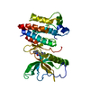
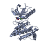
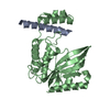
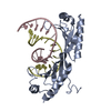
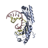
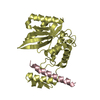
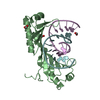
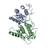
THREE-DIMENSIONAL STRUCTURE OF A COMPLEX BETWEEN THE DEATH DOMAINS OF PELLE AND TUBE
Components
DEATH DOMAIN OF PELLE
DEATH DOMAIN OF TUBE
Keywords
APOPTOSIS / SIX-HELIX BUNDLE / LINEAR ARRAY OF DEATH DOMAINS / PLASTIC INTERFACES
Function / homology
Function and homology information
MyD88 dependent cascade initiated on endosome / determination of dorsal/ventral asymmetry / hemocyte proliferation / Formation of the trans-membrane 'signalling complex' / Adaptor protein complex binds to TL receptor at the plasma membrane / DL and DIF homodimers bind to TUB and phosphorylated PLL in the TL receptor 'signalling complex' / DL and DIF homodimers complexed with CACT are all phosphorylated in the TL receptor 'signalling complex' / Activated PLL kinase is autophosphorylated in the TL receptor 'signalling complex' / Phosphorylated CACT, DL and DIF homodimers dissociate from the TL receptor 'signalling complex' / PLL kinase binds to TUB in the TL receptor 'signalling complex' ...MyD88 dependent cascade initiated on endosome / determination of dorsal/ventral asymmetry / hemocyte proliferation / Formation of the trans-membrane 'signalling complex' / Adaptor protein complex binds to TL receptor at the plasma membrane / DL and DIF homodimers bind to TUB and phosphorylated PLL in the TL receptor 'signalling complex' / DL and DIF homodimers complexed with CACT are all phosphorylated in the TL receptor 'signalling complex' / Activated PLL kinase is autophosphorylated in the TL receptor 'signalling complex' / Phosphorylated CACT, DL and DIF homodimers dissociate from the TL receptor 'signalling complex' / PLL kinase binds to TUB in the TL receptor 'signalling complex' / positive regulation of antifungal peptide production / TAK1-dependent IKK and NF-kappa-B activation / activated TAK1 mediates p38 MAPK activation / IRAK1 recruits IKK complex / IRAK2 mediated activation of TAK1 complex / TRAF6-mediated induction of TAK1 complex within TLR4 complex / IRAK1 recruits IKK complex upon TLR7/8 or 9 stimulation / IRAK2 mediated activation of TAK1 complex upon TLR7/8 or 9 stimulation / JNK (c-Jun kinases) phosphorylation and activation mediated by activated human TAK1 / zygotic specification of dorsal/ventral axis / Interleukin-1 signaling / positive regulation of hippo signaling / larval somatic muscle development / histone H3T3 kinase activity / antifungal innate immune response / Toll signaling pathway / response to fungus / positive regulation of ubiquitin-dependent protein catabolic process / dorsal/ventral pattern formation / protein tyrosine kinase binding / cytoplasmic side of plasma membrane / cytokine-mediated signaling pathway / positive regulation of protein catabolic process / mitotic cell cycle / cellular response to lipopolysaccharide / protein autophosphorylation / non-specific serine/threonine protein kinase / intracellular signal transduction / protein domain specific binding / protein phosphorylation / protein serine kinase activity / innate immune response / protein serine/threonine kinase activity / apoptotic process / ATP binding / nucleus / plasma membrane / cytosol / cytoplasm Similarity search - Function
Tube, Death domain / Tube Death domain / Pelle, death domain / Death Domain, Fas / Death Domain, Fas / Death domain profile. / DEATH domain, found in proteins involved in cell death (apoptosis). / Death domain / Death domain / Death-like domain superfamily ...Tube, Death domain / Tube Death domain / Pelle, death domain / Death Domain, Fas / Death Domain, Fas / Death domain profile. / DEATH domain, found in proteins involved in cell death (apoptosis). / Death domain / Death domain / Death-like domain superfamily / Protein tyrosine and serine/threonine kinase / Serine-threonine/tyrosine-protein kinase, catalytic domain / Serine/threonine-protein kinase, active site / Serine/Threonine protein kinases active-site signature. / Serine/Threonine protein kinases, catalytic domain / Protein kinase, ATP binding site / Protein kinases ATP-binding region signature. / Protein kinase domain profile. / Protein kinase domain / Protein kinase-like domain superfamily / Orthogonal Bundle / Mainly Alpha Similarity search - Domain/homology
Mass: 12446.343 Da / Num. of mol.: 2 Source method: isolated from a genetically manipulated source Source: (gene. exp.) Drosophila melanogaster (fruit fly) / Production host: Escherichia coli (E. coli) Keywords: THE FIRST FOUR RESIDUES OBSERVED IN THE STRUCTURE ORIGINATE FROM CLONING ARTEFACT. References: UniProt: Q05652
#2: Protein
DEATHDOMAINOFTUBE
Mass: 17084.508 Da / Num. of mol.: 2 Source method: isolated from a genetically manipulated source Source: (gene. exp.) Drosophila melanogaster (fruit fly) / Production host: Escherichia coli (E. coli) / References: UniProt: P22812
In the structure databanks used in Yorodumi, some data are registered as the other names, "COVID-19 virus" and "2019-nCoV". Here are the details of the virus and the list of structure data.
Jan 31, 2019. EMDB accession codes are about to change! (news from PDBe EMDB page)
EMDB accession codes are about to change! (news from PDBe EMDB page)
The allocation of 4 digits for EMDB accession codes will soon come to an end. Whilst these codes will remain in use, new EMDB accession codes will include an additional digit and will expand incrementally as the available range of codes is exhausted. The current 4-digit format prefixed with “EMD-” (i.e. EMD-XXXX) will advance to a 5-digit format (i.e. EMD-XXXXX), and so on. It is currently estimated that the 4-digit codes will be depleted around Spring 2019, at which point the 5-digit format will come into force.
The EM Navigator/Yorodumi systems omit the EMD- prefix.
Related info.:Q: What is EMD? / ID/Accession-code notation in Yorodumi/EM Navigator
Yorodumi is a browser for structure data from EMDB, PDB, SASBDB, etc.
This page is also the successor to EM Navigator detail page, and also detail information page/front-end page for Omokage search.
The word "yorodu" (or yorozu) is an old Japanese word meaning "ten thousand". "mi" (miru) is to see.
Related info.:EMDB / PDB / SASBDB / Comparison of 3 databanks / Yorodumi Search / Aug 31, 2016. New EM Navigator & Yorodumi / Yorodumi Papers / Jmol/JSmol / Function and homology information / Changes in new EM Navigator and Yorodumi
 Movie
Movie Controller

 Yorodumi
 Open data
 Basic information
 Components
 Keywords
 APOPTOSIS / SIX-HELIX BUNDLE / LINEAR ARRAY OF DEATH DOMAINS / PLASTIC INTERFACES
 Function and homology information
 Authors
 Citation
 Structure visualization
 Downloads & links
 Other downloads


 PDBj
PDBj


 Assembly
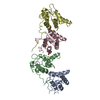


 Mass: 238.305 Da / Num. of mol.: 1 / Source method: obtained synthetically / Formula: C8H18N2O4S / Comment: pH buffer*YM
 Mass: 18.015 Da / Num. of mol.: 266 / Source method: isolated from a natural source / Formula: H2O
 Sample preparation
 / Beamline: 5.0.2 / Wavelength: 0.981
  Processing