 Movie
Movie Controller


+ Open data

- Basic information
| Entry | Database: PDB / ID: 1czw | ||||||
|---|---|---|---|---|---|---|---|
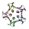
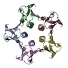
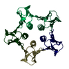
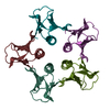
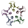
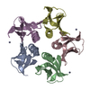
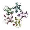
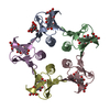
| Title | STRUCTURE OF THE W34A MUTANT OF SHIGA-LIKE TOXIN I B SUBUNIT | ||||||
 Components Components | SHIGA TOXIN B-CHAIN | ||||||
 Keywords Keywords |  TOXIN / BACTERIAL TOXIN / SUGAR RECEPTOR BINDING DOMAIN / PROTEIN-CARBOHYDRATE RECOGNITION / OB-FOLD TOXIN / BACTERIAL TOXIN / SUGAR RECEPTOR BINDING DOMAIN / PROTEIN-CARBOHYDRATE RECOGNITION / OB-FOLD | ||||||
| Function / homology |  Function and homology information Function and homology informationmodulation of host virulence by virus / hemolysis by symbiont of host erythrocytes / toxin activity / extracellular regionSimilarity search - Function | ||||||
| Biological species |  Phage h30 (virus) Phage h30 (virus) | ||||||
| Method | X-RAY DIFFRACTION / Resolution: 2.5 Å | ||||||
 Authors Authors | Ling, H. / Boodhoo, A. / Brunton, J.L. / Read, R.J. | ||||||
 Citation Citation | Journal: To be Published Title: A native structure of the W34A mutant at 2.5 A Authors: Ling, H. / Boodhoo, A. / Brunton, J.L. / Read, R.J. | ||||||
| History |
|
- Structure visualization
| Structure viewer | Molecule: MolmilJmol/JSmol |
|---|
- Downloads & links
-Download
| PDBx/mmCIF format | 1czw.cif.gz | 138.1 KB | Display | PDBx/mmCIF format |
|---|---|---|---|---|
| PDB format | pdb1czw.ent.gz | 115.9 KB | Display | PDB format |
| PDBx/mmJSON format | 1czw.json.gz | Tree view | PDBx/mmJSON format | |
| Others |  Other downloads Other downloads |
-Validation report
| Arichive directory | https://data.pdbj.org/pub/pdb/validation_reports/cz/1czwftp://data.pdbj.org/pub/pdb/validation_reports/cz/1czw | HTTPS FTP |
|---|
-Related structure data
| Related structure data | |
|---|---|
| Similar structure data |


-Links
 PDBj
PDBj
- Assembly
| Deposited unit | 
| ||||||||
|---|---|---|---|---|---|---|---|---|---|
| 1 | 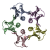
| ||||||||
| 2 | 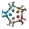
| ||||||||
| Unit cell |
|
-Components
| #1: Protein | Mass: 7583.501 Da / Num. of mol.: 10 / Fragment: SHIGA-LIKE TOXIN I BINDING DOMAIN / Mutation: W34A Source method: isolated from a genetically manipulated source Source: (gene. exp.) Phage h30 (virus) / Production host:  Escherichia coli (E. coli) / References: UniProt: P69178, UniProt: P69179*PLUS Escherichia coli (E. coli) / References: UniProt: P69178, UniProt: P69179*PLUS#2: Water | ChemComp-HOH / | Water Mass: 18.015 Da / Num. of mol.: 264 / Source method: isolated from a natural source / Formula: H2O Mass: 18.015 Da / Num. of mol.: 264 / Source method: isolated from a natural source / Formula: H2O |
|---|
-Experimental details
-Experiment
| Experiment | Method: X-RAY DIFFRACTION / Number of used crystals: 1 |
|---|
- Sample preparation
| Crystal | Density Matthews: 2.21 Å3/Da / Density % sol: 44.46 % |
|---|---|
| Crystal grow | Temperature: 298 K / Method: vapor diffusion, hanging drop / pH: 8 Details: 15% PEG 4000 0.2 M MGCL2 5% DIOXANE, pH 8.0, VAPOR DIFFUSION, HANGING DROP, temperature 298.0K |
-Data collection
| Diffraction | Mean temperature: 298 K |
|---|---|
| Diffraction source | Source: ROTATING ANODE / Type: SIEMENS / Wavelength: 1.5418 |
| Detector | Type: SIEMENS / Detector: AREA DETECTOR / Date: Apr 22, 1995 |
| Radiation | Protocol: SINGLE WAVELENGTH / Monochromatic (M) / Laue (L): M / Scattering type: x-ray |
| Radiation wavelength | Wavelength: 1.5418 Å / Relative weight: 1 |
| Reflection | Resolution: 2.5→48.6 Å / Num. all: 24009 / Num. obs: 23240 / % possible obs: 96.8 % / Observed criterion σ(F): 0 / Observed criterion σ(I): 0 / Redundancy: 6.7 % / Biso Wilson estimate: 38.53 Å2 / Rmerge(I) obs: 0.127 / Net I/σ(I): 11.5 |
| Reflection shell | Resolution: 2.5→2.54 Å / Redundancy: 2.5 % / Rmerge(I) obs: 0.365 / % possible all: 53.3 |
- Processing
| Software |
| ||||||||||||||||||||||||||||||||||||||||||||||||||||||||||||
|---|---|---|---|---|---|---|---|---|---|---|---|---|---|---|---|---|---|---|---|---|---|---|---|---|---|---|---|---|---|---|---|---|---|---|---|---|---|---|---|---|---|---|---|---|---|---|---|---|---|---|---|---|---|---|---|---|---|---|---|---|---|
| Refinement | Resolution: 2.5→48.6 Å / σ(F): 0 / σ(I): 0 / Stereochemistry target values: ENGH & HUBER / Details: MAXIMUM LIKELIHOOD F TARGET, WITH NCS RESTRAINTS
| ||||||||||||||||||||||||||||||||||||||||||||||||||||||||||||
| Displacement parameters |
| ||||||||||||||||||||||||||||||||||||||||||||||||||||||||||||
| Refinement step | Cycle: LAST / Resolution: 2.5→48.6 Å
| ||||||||||||||||||||||||||||||||||||||||||||||||||||||||||||
| Refine LS restraints |
|
