 Movie
Movie Controller


+ Open data

- Basic information
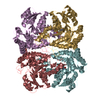
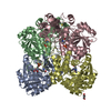
| Entry | Database: PDB / ID: 1cs1 | ||||||
|---|---|---|---|---|---|---|---|
| Title | CYSTATHIONINE GAMMA-SYNTHASE (CGS) FROM ESCHERICHIA COLI | ||||||
 Components Components | PROTEIN (CYSTATHIONINE GAMMA-SYNTHASE) | ||||||
 Keywords Keywords |  LYASE / LLP-DEPENDENT ENZYMES / METHIONINE BIOSYNTHESIS LYASE / LLP-DEPENDENT ENZYMES / METHIONINE BIOSYNTHESIS | ||||||
| Function / homology |  Function and homology informationcystathionine gamma-synthase / cystathionine gamma-synthase activity (acts on O-phosphohomoserine) / carbon-sulfur lyase activity / cystathionine gamma-synthase activity / cystathionine gamma-lyase activity / cysteine biosynthetic process via cystathionine / methionine biosynthetic process / transsulfuration / pyridoxal phosphate binding / cytoplasm Function and homology informationcystathionine gamma-synthase / cystathionine gamma-synthase activity (acts on O-phosphohomoserine) / carbon-sulfur lyase activity / cystathionine gamma-synthase activity / cystathionine gamma-lyase activity / cysteine biosynthetic process via cystathionine / methionine biosynthetic process / transsulfuration / pyridoxal phosphate binding / cytoplasmSimilarity search - Function | ||||||
| Biological species |  Escherichia coli (E. coli) Escherichia coli (E. coli) | ||||||
| Method | X-RAY DIFFRACTION / SYNCHROTRON / MOLECULAR REPLACEMENT / Resolution: 1.5 Å | ||||||
 Authors Authors | Clausen, T. / Messerschmidt, A. | ||||||
 Citation Citation | Journal: EMBO J. / Year: 1998 Title: Crystal structure of Escherichia coli cystathionine gamma-synthase at 1.5 A resolution. Authors: Clausen, T. / Huber, R. / Prade, L. / Wahl, M.C. / Messerschmidt, A. #1: Journal: FEBS Lett. / Year: 1997Title: Cloning, Purification, Crystallization and Preliminary Xray-Diffraction Analysis of Cystathionine Gamma-Synthase from Escherichia Coli Authors: Wahl, M.C. / Huber, R. / Prade, L. / Marinkovic, S. / Messerschmidt, A. / Clausen, T. | ||||||
| History |
|
- Structure visualization
| Structure viewer | Molecule: MolmilJmol/JSmol |
|---|
- Downloads & links
-Download
| PDBx/mmCIF format | 1cs1.cif.gz | 325.3 KB | Display | PDBx/mmCIF format |
|---|---|---|---|---|
| PDB format | pdb1cs1.ent.gz | 262.3 KB | Display | PDB format |
| PDBx/mmJSON format | 1cs1.json.gz | Tree view | PDBx/mmJSON format | |
| Others |  Other downloads Other downloads |
-Validation report
| Arichive directory | https://data.pdbj.org/pub/pdb/validation_reports/cs/1cs1ftp://data.pdbj.org/pub/pdb/validation_reports/cs/1cs1 | HTTPS FTP |
|---|
-Related structure data
| Related structure data | 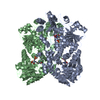 1cl1S S: Starting model for refinement |
|---|---|
| Similar structure data |


-Links
 PDBj
PDBj- Assembly
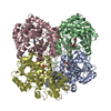
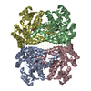
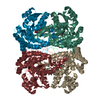
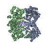
| Deposited unit | 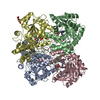
| ||||||||
|---|---|---|---|---|---|---|---|---|---|
| 1 |
| ||||||||
| Unit cell |
| ||||||||
| Components on special symmetry positions |
|
-Components
| #1: Protein | Mass: 41825.414 Da / Num. of mol.: 4 Source method: isolated from a genetically manipulated source Details: PLP BOUND AS COFACTOR TO LYS 198 / Source: (gene. exp.) Escherichia coli (E. coli) / Strain: DH5 / Gene: METB / Species (production host): Escherichia coli / Production host: Escherichia coli BL21(DE3) (bacteria) / Strain (production host): BL21/DE3 / References: UniProt: P00935, EC: 4.2.99.9#2: Chemical | ChemComp-DHD / |   Mass: 160.082 Da / Num. of mol.: 1 / Source method: obtained synthetically / Formula: C5H4O6 Mass: 160.082 Da / Num. of mol.: 1 / Source method: obtained synthetically / Formula: C5H4O6#3: Water | ChemComp-HOH / | Water Mass: 18.015 Da / Num. of mol.: 1301 / Source method: isolated from a natural source / Formula: H2O Mass: 18.015 Da / Num. of mol.: 1301 / Source method: isolated from a natural source / Formula: H2O |
|---|
-Experimental details
-Experiment
| Experiment | Method: X-RAY DIFFRACTION / Number of used crystals: 1 |
|---|
- Sample preparation
| Crystal | Density Matthews: 2.3 Å3/Da / Density % sol: 55 % | ||||||||||||||||||||||||||||||||||||||||
|---|---|---|---|---|---|---|---|---|---|---|---|---|---|---|---|---|---|---|---|---|---|---|---|---|---|---|---|---|---|---|---|---|---|---|---|---|---|---|---|---|---|
| Crystal grow | pH: 7.45 / Details: 17% PEG400, 10% MPD, 0.1 M MES/NAOH (PH 7.45) | ||||||||||||||||||||||||||||||||||||||||
| Crystal | *PLUS | ||||||||||||||||||||||||||||||||||||||||
| Crystal grow | *PLUS pH: 7 / Method: vapor diffusion, sitting drop / Details: Wahl, M.C., (1997) FEBS Lett., 414, 492. | ||||||||||||||||||||||||||||||||||||||||
| Components of the solutions | *PLUS
|
-Data collection
| Diffraction | Mean temperature: 100 K |
|---|---|
| Diffraction source | Source: SYNCHROTRON / Site: MPG/DESY, HAMBURG  / Beamline: BW6 / Wavelength: 1.1 / Beamline: BW6 / Wavelength: 1.1 |
| Detector | Type: MARRESEARCH / Detector: IMAGE PLATE / Date: Sep 15, 1997 / Details: MIRRORS |
| Radiation | Monochromator: SINGLE CRYSTAL / Protocol: SINGLE WAVELENGTH / Monochromatic (M) / Laue (L): M / Scattering type: x-ray |
| Radiation wavelength | Wavelength: 1.1 Å / Relative weight: 1 |
| Reflection | Resolution: 1.5→8 Å / Num. obs: 200884 / % possible obs: 86.9 % / Observed criterion σ(I): 2 / Redundancy: 2.7 % / Biso Wilson estimate: 26.3 Å2 / Rmerge(I) obs: 0.066 / Rsym value: 0.058 / Net I/σ(I): 23.4 |
| Reflection shell | Resolution: 1.5→1.55 Å / Redundancy: 2.9 % / Rmerge(I) obs: 0.205 / Mean I/σ(I) obs: 7.2 / Rsym value: 0.214 / % possible all: 70.1 |
| Reflection shell | *PLUS % possible obs: 70.1 % / Num. unique obs: 16087 |
- Processing
| Software |
| ||||||||||||||||||||||||||||||||||||||||||||||||||||||||||||
|---|---|---|---|---|---|---|---|---|---|---|---|---|---|---|---|---|---|---|---|---|---|---|---|---|---|---|---|---|---|---|---|---|---|---|---|---|---|---|---|---|---|---|---|---|---|---|---|---|---|---|---|---|---|---|---|---|---|---|---|---|---|
| Refinement | Method to determine structure: MOLECULAR REPLACEMENT Starting model: PDB ENTRY 1CL1 Resolution: 1.5→8 Å / Rfactor Rfree error: 0.017 / Data cutoff high absF: 1000000 / Data cutoff low absF: 0.001 / Cross valid method: THROUGHOUT / σ(F): 2
| ||||||||||||||||||||||||||||||||||||||||||||||||||||||||||||
| Displacement parameters | Biso mean: 30.8 Å2 | ||||||||||||||||||||||||||||||||||||||||||||||||||||||||||||
| Refine analyze | Luzzati coordinate error obs: 0.17 Å / Luzzati d res low obs: 8 Å | ||||||||||||||||||||||||||||||||||||||||||||||||||||||||||||
| Refinement step | Cycle: LAST / Resolution: 1.5→8 Å
| ||||||||||||||||||||||||||||||||||||||||||||||||||||||||||||
| Refine LS restraints |
| ||||||||||||||||||||||||||||||||||||||||||||||||||||||||||||
| Xplor file | Serial no: 1 / Param file: PARAM19X.PRO / Topol file: TOPH19X.PRO | ||||||||||||||||||||||||||||||||||||||||||||||||||||||||||||
| Software | *PLUS Name: X-PLOR / Version: 3.1 / Classification: refinement | ||||||||||||||||||||||||||||||||||||||||||||||||||||||||||||
| Refinement | *PLUS Highest resolution: 1.5 Å / Lowest resolution: 8 Å / σ(F): 2 / % reflection Rfree: 10 % / Rfactor obs: 0.2 / Rfactor Rwork: 0.2 | ||||||||||||||||||||||||||||||||||||||||||||||||||||||||||||
| Solvent computation | *PLUS | ||||||||||||||||||||||||||||||||||||||||||||||||||||||||||||
| Displacement parameters | *PLUS Biso mean: 30.8 Å2 | ||||||||||||||||||||||||||||||||||||||||||||||||||||||||||||
| Refine LS restraints | *PLUS
|
