 Movie
Movie Controller


+ Open data

- Basic information
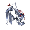
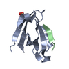
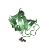
| Entry | Database: PDB / ID: 1cm2 | ||||||
|---|---|---|---|---|---|---|---|
| Title | STRUCTURE OF HIS15ASP HPR AFTER HYDROLYSIS OF RINGED SPECIES. | ||||||
 Components Components | HISTIDINE-CONTAINING PROTEIN | ||||||
 Keywords Keywords |  TRANSFERASE / PHOSPHOTRANSFERASE / SUCCINIMIDE / ISOIMIDE TRANSFERASE / PHOSPHOTRANSFERASE / SUCCINIMIDE / ISOIMIDE | ||||||
| Function / homology |  Function and homology information Function and homology informationphosphotransferase activity, nitrogenous group as acceptor / regulation of carbon utilization / antisigma factor binding / positive regulation of glycogen catabolic process / phosphoenolpyruvate-dependent sugar phosphotransferase system / enzyme inhibitor activity / enzyme activator activity / enzyme regulator activity / cytosolSimilarity search - Function | ||||||
| Biological species |  Escherichia coli (E. coli) Escherichia coli (E. coli) | ||||||
| Method | X-RAY DIFFRACTION / SYNCHROTRON / MOLECULAR REPLACEMENT / Resolution: 1.8 Å | ||||||
 Authors Authors | Napper, S. / Delbaere, L.T.J. / Waygood, E.B. | ||||||
 Citation Citation | Journal: J.Biol.Chem. / Year: 1999 Title: The aspartyl replacement of the active site histidine in histidine-containing protein, HPr, of the Escherichia coli Phosphoenolpyruvate:Sugar phosphotransferase system can accept and donate a ...Title: The aspartyl replacement of the active site histidine in histidine-containing protein, HPr, of the Escherichia coli Phosphoenolpyruvate:Sugar phosphotransferase system can accept and donate a phosphoryl group. Spontaneous dephosphorylation of acyl-phosphate autocatalyzes an internal cyclization Authors: Napper, S. / Delbaere, L.T. / Waygood, E.B. | ||||||
| History |
|
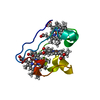
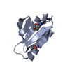
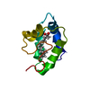
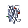
- Structure visualization
| Structure viewer | Molecule: MolmilJmol/JSmol |
|---|
- Downloads & links
-Download
| PDBx/mmCIF format | 1cm2.cif.gz | 32.2 KB | Display | PDBx/mmCIF format |
|---|---|---|---|---|
| PDB format | pdb1cm2.ent.gz | 22 KB | Display | PDB format |
| PDBx/mmJSON format | 1cm2.json.gz | Tree view | PDBx/mmJSON format | |
| Others |  Other downloads Other downloads |
-Validation report
| Arichive directory | https://data.pdbj.org/pub/pdb/validation_reports/cm/1cm2ftp://data.pdbj.org/pub/pdb/validation_reports/cm/1cm2 | HTTPS FTP |
|---|
-Related structure data


-Links
 PDBj
PDBj- Assembly
| Deposited unit | 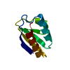
| ||||||||
|---|---|---|---|---|---|---|---|---|---|
| 1 |
| ||||||||
| Unit cell |
|
-Components
| #1: Protein | Mass: 9106.273 Da / Num. of mol.: 1 / Mutation: HIS15ASP Source method: isolated from a genetically manipulated source Details: THE STRUCTURE IS THAT OF HIS15ASP HPR WHICH HAS UNDERGONE HYDROLYSIS FROM A HIGH-PI RINGED SPECIES OF THE PROTEIN WHICH IS BELIEVED TO INVOLVE SUCCINIMIDE OR ISOIMIDE FORMATION. Source: (gene. exp.) Escherichia coli (E. coli) / Gene: PTSH / Plasmid: PUC19 / Cell line (production host): ESK108 / Gene (production host): PTSH / Production host: Escherichia coli (E. coli) / References: UniProt: P0AA04 |
|---|---|
| #2: Water | ChemComp-HOH / Water Mass: 18.015 Da / Num. of mol.: 48 / Source method: isolated from a natural source / Formula: H2O Mass: 18.015 Da / Num. of mol.: 48 / Source method: isolated from a natural source / Formula: H2O |
-Experimental details
-Experiment
| Experiment | Method: X-RAY DIFFRACTION / Number of used crystals: 1 |
|---|
- Sample preparation
| Crystal | Density Matthews: 1.73 Å3/Da / Density % sol: 28.75 % | |||||||||||||||
|---|---|---|---|---|---|---|---|---|---|---|---|---|---|---|---|---|
| Crystal grow | pH: 4.6 / Details: pH 4.6 | |||||||||||||||
| Crystal grow | *PLUS Temperature: 14 ℃ / pH: 4.4 / Method: vapor diffusion, hanging drop | |||||||||||||||
| Components of the solutions | *PLUS
|
-Data collection
| Diffraction | Mean temperature: 273 K |
|---|---|
| Diffraction source | Source: SYNCHROTRON / Site: Photon Factory  / Type: PHOTON FACTORY / Wavelength: 1 / Type: PHOTON FACTORY / Wavelength: 1 |
| Detector | Type: WEISSENBERG |
| Radiation | Protocol: SINGLE WAVELENGTH / Monochromatic (M) / Laue (L): M / Scattering type: x-ray |
| Radiation wavelength | Wavelength: 1 Å / Relative weight: 1 |
| Reflection | Resolution: 1.8→10 Å / Num. obs: 6570 / % possible obs: 97.5 % / Observed criterion σ(I): 0 / Redundancy: 2.1 % / Rmerge(I) obs: 0.07 / Rsym value: 9.7 |
- Processing
| Software |
| ||||||||||||||||||||||||||||||||||||||||||||||||||||||||||||
|---|---|---|---|---|---|---|---|---|---|---|---|---|---|---|---|---|---|---|---|---|---|---|---|---|---|---|---|---|---|---|---|---|---|---|---|---|---|---|---|---|---|---|---|---|---|---|---|---|---|---|---|---|---|---|---|---|---|---|---|---|---|
| Refinement | Method to determine structure: MOLECULAR REPLACEMENT Starting model: WILD TYPE E. COLI HPR Resolution: 1.8→10 Å / σ(F): 0
| ||||||||||||||||||||||||||||||||||||||||||||||||||||||||||||
| Refinement step | Cycle: LAST / Resolution: 1.8→10 Å
| ||||||||||||||||||||||||||||||||||||||||||||||||||||||||||||
| Refine LS restraints |
| ||||||||||||||||||||||||||||||||||||||||||||||||||||||||||||
| Software | *PLUS Name: X-PLOR / Version: 3.2 / Classification: refinement | ||||||||||||||||||||||||||||||||||||||||||||||||||||||||||||
| Refinement | *PLUS Highest resolution: 1.8 Å / Lowest resolution: 10 Å / σ(F): 0 / Rfactor obs: 0.2 / Rfactor Rwork: 0.2 / Num. reflection obs: 5670 | ||||||||||||||||||||||||||||||||||||||||||||||||||||||||||||
| Solvent computation | *PLUS | ||||||||||||||||||||||||||||||||||||||||||||||||||||||||||||
| Displacement parameters | *PLUS | ||||||||||||||||||||||||||||||||||||||||||||||||||||||||||||
| Refine LS restraints | *PLUS Type: x_angle_deg / Dev ideal: 1.4 |
