 Movie
Movie Controller


+ Open data

- Basic information
| Entry | Database: PDB / ID: 1cgo | ||||||||||||
|---|---|---|---|---|---|---|---|---|---|---|---|---|---|
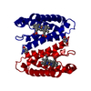
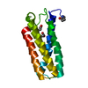
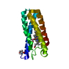
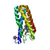
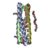
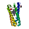
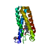
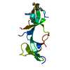
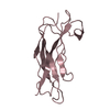
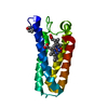
| Title | CYTOCHROME C' | ||||||||||||
 Components Components | CYTOCHROME C | ||||||||||||
 Keywords Keywords | ELECTRON TRANSPORT (CYTOCHROME) | ||||||||||||
| Function / homology |  Function and homology informationelectron transfer activity / periplasmic space / iron ion binding / heme binding Function and homology informationelectron transfer activity / periplasmic space / iron ion binding / heme bindingSimilarity search - Function | ||||||||||||
| Biological species |  Alcaligenes sp. (bacteria) Alcaligenes sp. (bacteria) | ||||||||||||
| Method | X-RAY DIFFRACTION / SYNCHROTRON / Resolution: 1.8 Å | ||||||||||||
 Authors Authors | Dobbs, A.J. / Faber, H.R. / Anderson, B.F. / Baker, E.N. | ||||||||||||
 Citation Citation | Journal: Acta Crystallogr.,Sect.D / Year: 1996 Title: Three-dimensional structure of cytochrome c' from two Alcaligenes species and the implications for four-helix bundle structures. Authors: Dobbs, A.J. / Anderson, B.F. / Faber, H.R. / Baker, E.N. #1: Journal: J.Mol.Biol. / Year: 1993Title: Atomic Structure of a Cytochrome C' with an Unusual Ligand-Controlled Dimer Dissociation at 1.8 Angstroms Resolution Authors: Ren, Z. / Meyer, T.E. / Mcree, D.E. #2: Journal: J.Biochem.(Tokyo) / Year: 1992Title: Three-Dimensional Structure of Ferricytochrome C' from Rhodospirillum Rubrum at 2.8 Angstroms Resolution Authors: Yasui, M. / Harada, S. / Kai, Y. / Kasai, N. / Kusunoki, M. / Matsura, Y. #3: Journal: J.Mol.Biol. / Year: 1985Title: Structure of Ferricytochrome C' from Rhodospirillum Molischianum at 1.67 Angstroms Authors: Finzel, B.C. / Weber, P.C. / Hardman, K.D. / Salemme, F.R. #4: Journal: J.Mol.Biol. / Year: 1981Title: Crystallographic Structure of Rhodospirillum Molischianum Ferricytochrome C' at 2.5 Angstroms Resolution Authors: Weber, P.C. / Howard, A. / Xoung, N.H. / Salemme, F.R. #5: Journal: Biochem.J. / Year: 1973Title: The Amino Acid Sequence of Cytochrome C' from Alcaligenes Sp. N.C.I.B. 11015 Authors: Ambler, R.P. | ||||||||||||
| History |
|
- Structure visualization
| Structure viewer | Molecule: MolmilJmol/JSmol |
|---|
- Downloads & links
-Download
| PDBx/mmCIF format | 1cgo.cif.gz | 35.6 KB | Display | PDBx/mmCIF format |
|---|---|---|---|---|
| PDB format | pdb1cgo.ent.gz | 27 KB | Display | PDB format |
| PDBx/mmJSON format | 1cgo.json.gz | Tree view | PDBx/mmJSON format | |
| Others |  Other downloads Other downloads |
-Validation report
| Arichive directory | https://data.pdbj.org/pub/pdb/validation_reports/cg/1cgoftp://data.pdbj.org/pub/pdb/validation_reports/cg/1cgo | HTTPS FTP |
|---|
-Related structure data

-Links
 PDBj
PDBj


- Assembly
| Deposited unit | 
| ||||||||
|---|---|---|---|---|---|---|---|---|---|
| 1 | 
| ||||||||
| Unit cell |
| ||||||||
| Details | SYMMETRY THE CRYSTALLOGRAPHIC SYMMETRY TRANSFORMATIONS PRESENTED BELOW GENERATE THE SUBUNITS OF THE POLYMERIC MOLECULE. APPLIED TO RESIDUES: PCA 1 .. HEM 128 SYMMETRY OPERATION TO GENERATE THE SECOND MOLECULE OF THE DIMERIC PARTICLE SYMMETRY1 1 -1.000000 0.000000 0.000000 0.00000 SYMMETRY2 1 0.000000 1.000000 0.000000 0.00000 SYMMETRY3 1 0.000000 0.000000 -1.000000 90.55000 |
-Components
| #1: Protein | Mass: 13645.469 Da / Num. of mol.: 1 Source method: isolated from a genetically manipulated source Source: (gene. exp.) Alcaligenes sp. (bacteria) / References: UniProt: P00138 |
|---|---|
| #2: Chemical | ChemComp-HEC / Heme C  Mass: 618.503 Da / Num. of mol.: 1 / Source method: obtained synthetically / Formula: C34H34FeN4O4 Mass: 618.503 Da / Num. of mol.: 1 / Source method: obtained synthetically / Formula: C34H34FeN4O4 |
| #3: Water | ChemComp-HOH / Water Mass: 18.015 Da / Num. of mol.: 89 / Source method: isolated from a natural source / Formula: H2O Mass: 18.015 Da / Num. of mol.: 89 / Source method: isolated from a natural source / Formula: H2O |
-Experimental details
-Experiment
| Experiment | Method: X-RAY DIFFRACTION |
|---|
- Sample preparation
| Crystal | Density Matthews: 2.83 Å3/Da / Density % sol: 56.59 % |
|---|
-Data collection
| Diffraction source | Source: SYNCHROTRON / Site: Photon Factory  / Beamline: BL-6A / Wavelength: 1.04 / Beamline: BL-6A / Wavelength: 1.04 |
|---|---|
| Detector | Detector: IMAGE PLATE / Date: Nov 28, 1990 |
| Radiation | Monochromatic (M) / Laue (L): M / Scattering type: x-ray |
| Radiation wavelength | Wavelength: 1.04 Å / Relative weight: 1 |
| Reflection | Redundancy: 5.2 % / Rmerge(I) obs: 0.075 |
- Processing
| Software |
| ||||||||||||
|---|---|---|---|---|---|---|---|---|---|---|---|---|---|
| Refinement | Resolution: 1.8→20 Å / σ(F): 0 /
| ||||||||||||
| Refinement step | Cycle: LAST / Resolution: 1.8→20 Å
|
