 Movie
Movie Controller

[English] 日本語
 Yorodumi
- PDB-1cbu: ADENOSYLCOBINAMIDE KINASE/ADENOSYLCOBINAMIDE PHOSPHATE GUANYLYLTR... -
+ Open data

- Basic information
| Entry | Database: PDB / ID: 1cbu | ||||||
|---|---|---|---|---|---|---|---|
| Title | ADENOSYLCOBINAMIDE KINASE/ADENOSYLCOBINAMIDE PHOSPHATE GUANYLYLTRANSFERASE (COBU) FROM SALMONELLA TYPHIMURIUM | ||||||
 Components Components | ADENOSYLCOBINAMIDE KINASE/ADENOSYLCOBINAMIDE PHOSPHATE GUANYLYLTRANSFERASE | ||||||
 Keywords Keywords |  COBALAMIN BIOSYNTHESIS / ADENOSYLCOBINAMIDE / KINASE / ATPASE / GUANYLYLTRANSFERASE / SALMONELLA TYPHIMURIUM / TRANSFERASE / COENZYME B12 COBALAMIN BIOSYNTHESIS / ADENOSYLCOBINAMIDE / KINASE / ATPASE / GUANYLYLTRANSFERASE / SALMONELLA TYPHIMURIUM / TRANSFERASE / COENZYME B12 | ||||||
| Function / homology |  Function and homology informationadenosylcobinamide kinase / adenosylcobinamide kinase (GTP-specific) activity / adenosylcobinamide kinase (ATP-specific) activity / adenosylcobinamide-phosphate guanylyltransferase / cobinamide phosphate guanylyltransferase activity / cobalamin biosynthetic process / phosphorylation / GTP binding / ATP binding Function and homology informationadenosylcobinamide kinase / adenosylcobinamide kinase (GTP-specific) activity / adenosylcobinamide kinase (ATP-specific) activity / adenosylcobinamide-phosphate guanylyltransferase / cobinamide phosphate guanylyltransferase activity / cobalamin biosynthetic process / phosphorylation / GTP binding / ATP bindingSimilarity search - Function | ||||||
| Biological species |  Salmonella typhimurium (bacteria) Salmonella typhimurium (bacteria) | ||||||
| Method | X-RAY DIFFRACTION / MIR / Resolution: 2.3 Å | ||||||
 Authors Authors | Thompson, T.B. / Thomas, M.G. / Escalante-Semerena, J.C. / Rayment, I. | ||||||
 Citation Citation | Journal: Biochemistry / Year: 1998 Title: Three-dimensional structure of adenosylcobinamide kinase/adenosylcobinamide phosphate guanylyltransferase from Salmonella typhimurium determined to 2.3 A resolution,. Authors: Thompson, T.B. / Thomas, M.G. / Escalante-Semerena, J.C. / Rayment, I. #1: Journal: J.Biol.Chem. / Year: 1995Title: Purification and Characterization of the Bifunctional Cobu Enzyme of Salmonella Typhimurium Lt2. Evidence for a Cobu-Gmp Intermediate Authors: O'Toole, G.A. / Escalante-Semerena, J.C. | ||||||
| History |
|
- Structure visualization
| Structure viewer | Molecule: MolmilJmol/JSmol |
|---|
- Downloads & links
-Download
| PDBx/mmCIF format | 1cbu.cif.gz | 118 KB | Display | PDBx/mmCIF format |
|---|---|---|---|---|
| PDB format | pdb1cbu.ent.gz | 92.2 KB | Display | PDB format |
| PDBx/mmJSON format | 1cbu.json.gz | Tree view | PDBx/mmJSON format | |
| Others |  Other downloads Other downloads |
-Validation report
| Arichive directory | https://data.pdbj.org/pub/pdb/validation_reports/cb/1cbuftp://data.pdbj.org/pub/pdb/validation_reports/cb/1cbu | HTTPS FTP |
|---|
-Related structure data
| Similar structure data |
|---|


-Links
 PDBj
PDBj- Assembly
| Deposited unit | 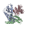
| ||||||||||||
|---|---|---|---|---|---|---|---|---|---|---|---|---|---|
| 1 |
| ||||||||||||
| Unit cell |
| ||||||||||||
| Noncrystallographic symmetry (NCS) | NCS oper:
|
-Components
| #1: Protein | Mass: 19794.711 Da / Num. of mol.: 3 Source method: isolated from a genetically manipulated source Source: (gene. exp.) Salmonella typhimurium (bacteria) / Production host: Escherichia coli (E. coli) / Strain (production host): LT2 / References: UniProt: Q05599#2: Chemical | Sulfate  Mass: 96.063 Da / Num. of mol.: 3 / Source method: obtained synthetically / Formula: SO4 Mass: 96.063 Da / Num. of mol.: 3 / Source method: obtained synthetically / Formula: SO4#3: Water | ChemComp-HOH / | Water Mass: 18.015 Da / Num. of mol.: 202 / Source method: isolated from a natural source / Formula: H2O Mass: 18.015 Da / Num. of mol.: 202 / Source method: isolated from a natural source / Formula: H2O |
|---|
-Experimental details
-Experiment
| Experiment | Method: X-RAY DIFFRACTION / Number of used crystals: 2 |
|---|
- Sample preparation
| Crystal | Density Matthews: 2.45 Å3/Da / Density % sol: 50 % | ||||||||||||||||||||||||||||||
|---|---|---|---|---|---|---|---|---|---|---|---|---|---|---|---|---|---|---|---|---|---|---|---|---|---|---|---|---|---|---|---|
| Crystal grow | Method: microbatch / pH: 5.5 Details: PROTEIN WAS CRYSTALLIZED BY MICRO BATCH FROM 6% PEG 3350, 200 MM NACL AND 50 MM SUCCINATE, PH 5.5 AT 4 C., micro batch | ||||||||||||||||||||||||||||||
| Crystal grow | *PLUS Temperature: 4 ℃ / Method: batch method | ||||||||||||||||||||||||||||||
| Components of the solutions | *PLUS
|
-Data collection
| Diffraction | Mean temperature: 263 K |
|---|---|
| Diffraction source | Source: ROTATING ANODE / Type: RIGAKU RUH2R / Wavelength: 1.5418 |
| Detector | Type: SIEMENS HI-STAR / Detector: AREA DETECTOR / Date: Sep 1, 1997 / Details: SIEMENS GOBEL FOCUSING OPTICS |
| Radiation | Monochromatic (M) / Laue (L): M / Scattering type: x-ray |
| Radiation wavelength | Wavelength: 1.5418 Å / Relative weight: 1 |
| Reflection | Highest resolution: 2.3 Å / Num. obs: 24136 / % possible obs: 88.5 % / Observed criterion σ(I): 0.33 / Redundancy: 3 % / Biso Wilson estimate: 46.9 Å2 / Rsym value: 0.043 / Net I/σ(I): 16 |
| Reflection shell | Resolution: 2.3→2.37 Å / Redundancy: 1.9 % / Mean I/σ(I) obs: 1.7 / Rsym value: 0.21 / % possible all: 64.4 |
| Reflection | *PLUS Rmerge(I) obs: 0.043 |
| Reflection shell | *PLUS % possible obs: 64.4 % / Rmerge(I) obs: 0.21 |
- Processing
| Software |
| ||||||||||||||||||||||||||||||||||||||||||||||||||
|---|---|---|---|---|---|---|---|---|---|---|---|---|---|---|---|---|---|---|---|---|---|---|---|---|---|---|---|---|---|---|---|---|---|---|---|---|---|---|---|---|---|---|---|---|---|---|---|---|---|---|---|
| Refinement | Method to determine structure: MIR / Resolution: 2.3→20 Å / σ(F): 0 / Stereochemistry target values: TNT PROTGEO
| ||||||||||||||||||||||||||||||||||||||||||||||||||
| Solvent computation | Solvent model: TNT / Bsol: 740 Å2 / ksol: 0.97 e/Å3 | ||||||||||||||||||||||||||||||||||||||||||||||||||
| Refinement step | Cycle: LAST / Resolution: 2.3→20 Å
| ||||||||||||||||||||||||||||||||||||||||||||||||||
| Refine LS restraints |
| ||||||||||||||||||||||||||||||||||||||||||||||||||
| Software | *PLUS Name: TNT / Version: 4C / Classification: refinement | ||||||||||||||||||||||||||||||||||||||||||||||||||
| Refine LS restraints | *PLUS
|
