 Movie
Movie Controller


+ Open data

- Basic information
| Entry | Database: PDB / ID: 1cb5 | ||||||
|---|---|---|---|---|---|---|---|
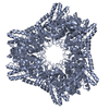
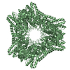
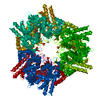
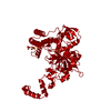
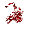
| Title | HUMAN BLEOMYCIN HYDROLASE. | ||||||
 Components Components | BLEOMYCIN HYDROLASE | ||||||
 Keywords Keywords | HYDROLASE / AMINOPEPTIDASE / CYSTEINE PROTEASE / SELF-COMPARTMENTALIZING / BLEOMYCIN | ||||||
| Function / homology |  Function and homology informationbleomycin hydrolase / cysteine-type aminopeptidase activity / homocysteine catabolic process / aminopeptidase activity / carboxypeptidase activity / cysteine-type peptidase activity / response to toxic substance / protein polyubiquitination / Antigen processing: Ubiquitination & Proteasome degradation / response to xenobiotic stimulus ...bleomycin hydrolase / cysteine-type aminopeptidase activity / homocysteine catabolic process / aminopeptidase activity / carboxypeptidase activity / cysteine-type peptidase activity / response to toxic substance / protein polyubiquitination / Antigen processing: Ubiquitination & Proteasome degradation / response to xenobiotic stimulus / cysteine-type endopeptidase activity / proteolysis / extracellular exosome / identical protein binding / nucleus / cytosol / cytoplasm Function and homology informationbleomycin hydrolase / cysteine-type aminopeptidase activity / homocysteine catabolic process / aminopeptidase activity / carboxypeptidase activity / cysteine-type peptidase activity / response to toxic substance / protein polyubiquitination / Antigen processing: Ubiquitination & Proteasome degradation / response to xenobiotic stimulus ...bleomycin hydrolase / cysteine-type aminopeptidase activity / homocysteine catabolic process / aminopeptidase activity / carboxypeptidase activity / cysteine-type peptidase activity / response to toxic substance / protein polyubiquitination / Antigen processing: Ubiquitination & Proteasome degradation / response to xenobiotic stimulus / cysteine-type endopeptidase activity / proteolysis / extracellular exosome / identical protein binding / nucleus / cytosol / cytoplasmSimilarity search - Function | ||||||
| Biological species |  Homo sapiens (human) Homo sapiens (human) | ||||||
| Method | X-RAY DIFFRACTION / SYNCHROTRON / MOLECULAR REPLACEMENT / Resolution: 2.59 Å | ||||||
 Authors Authors | O'Farrell, P.A. / Gonzalez, F. / Zheng, W. / Johnston, S.A. / Joshua-Tor, L. | ||||||
 Citation Citation | Journal: Structure Fold.Des. / Year: 1999 Title: Crystal structure of human bleomycin hydrolase, a self-compartmentalizing cysteine protease. Authors: O'Farrell, P.A. / Gonzalez, F. / Zheng, W. / Johnston, S.A. / Joshua-Tor, L. | ||||||
| History |
|
- Structure visualization
| Structure viewer | Molecule: MolmilJmol/JSmol |
|---|
- Downloads & links
-Download
| PDBx/mmCIF format | 1cb5.cif.gz | 270.6 KB | Display | PDBx/mmCIF format |
|---|---|---|---|---|
| PDB format | pdb1cb5.ent.gz | 222.2 KB | Display | PDB format |
| PDBx/mmJSON format | 1cb5.json.gz | Tree view | PDBx/mmJSON format | |
| Others |  Other downloads Other downloads |
-Validation report
| Arichive directory | https://data.pdbj.org/pub/pdb/validation_reports/cb/1cb5ftp://data.pdbj.org/pub/pdb/validation_reports/cb/1cb5 | HTTPS FTP |
|---|
-Related structure data
| Related structure data | 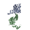 2cb5C  1gcbS S: Starting model for refinement C: citing same article ( |
|---|---|
| Similar structure data |


-Links
 PDBj
PDBj
- Assembly
| Deposited unit | 
| ||||||||||||
|---|---|---|---|---|---|---|---|---|---|---|---|---|---|
| 1 | 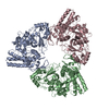
| ||||||||||||
| Unit cell |
| ||||||||||||
| Noncrystallographic symmetry (NCS) | NCS oper:
|
-Components
| #1: Protein | Mass: 52342.754 Da / Num. of mol.: 3 Source method: isolated from a genetically manipulated source Source: (gene. exp.) Homo sapiens (human) / Cellular location: CYTOPLASM / Plasmid: PKH260HUBH / Production host:  Escherichia coli (E. coli) / Strain (production host): BL21(DE3)/PLYSS Escherichia coli (E. coli) / Strain (production host): BL21(DE3)/PLYSSReferences: UniProt: Q13867, Hydrolases; Acting on peptide bonds (peptidases); Cysteine endopeptidases |
|---|
-Experimental details
-Experiment
| Experiment | Method: X-RAY DIFFRACTION / Number of used crystals: 1 |
|---|
- Sample preparation
| Crystal | Density Matthews: 2.65 Å3/Da / Density % sol: 53.59 % | ||||||||||||||||||||||||||||||||||||||||||
|---|---|---|---|---|---|---|---|---|---|---|---|---|---|---|---|---|---|---|---|---|---|---|---|---|---|---|---|---|---|---|---|---|---|---|---|---|---|---|---|---|---|---|---|
| Crystal grow | pH: 8 / Details: pH 8.00 | ||||||||||||||||||||||||||||||||||||||||||
| Crystal grow | *PLUS Temperature: 17 ℃ / pH: 8 / Method: vapor diffusion, hanging drop | ||||||||||||||||||||||||||||||||||||||||||
| Components of the solutions | *PLUS
|
-Data collection
| Diffraction | Mean temperature: 100 K |
|---|---|
| Diffraction source | Source: SYNCHROTRON / Site: NSLS  / Beamline: X26C / Wavelength: 1.126 / Beamline: X26C / Wavelength: 1.126 |
| Detector | Type: MARRESEARCH / Detector: IMAGE PLATE |
| Radiation | Protocol: SINGLE WAVELENGTH / Monochromatic (M) / Laue (L): M / Scattering type: x-ray |
| Radiation wavelength | Wavelength: 1.126 Å / Relative weight: 1 |
| Reflection | Resolution: 2.6→100 Å / Num. obs: 47154 / Observed criterion σ(I): 0 / Redundancy: 5.1 % / Biso Wilson estimate: 39.1 Å2 / Rsym value: 7.2 / Net I/σ(I): 18.8 |
| Reflection shell | Resolution: 2.59→2.68 Å / Mean I/σ(I) obs: 3.5 / Rsym value: 43.1 / % possible all: 94 |
| Reflection | *PLUS Highest resolution: 2.59 Å / Lowest resolution: 25 Å / % possible obs: 91.6 % / Observed criterion σ(I): 0 / Redundancy: 5.1 % / Num. measured all: 242494 / Rmerge(I) obs: 0.072 / Biso Wilson estimate: 39.1 Å2 |
| Reflection shell | *PLUS Highest resolution: 2.59 Å / Lowest resolution: 2.68 Å / % possible obs: 94 % / Rmerge(I) obs: 0.431 / Mean I/σ(I) obs: 3.5 |
- Processing
| Software |
| ||||||||||||||||||||||||||||||||||||||||||||||||||||||||||||
|---|---|---|---|---|---|---|---|---|---|---|---|---|---|---|---|---|---|---|---|---|---|---|---|---|---|---|---|---|---|---|---|---|---|---|---|---|---|---|---|---|---|---|---|---|---|---|---|---|---|---|---|---|---|---|---|---|---|---|---|---|---|
| Refinement | Method to determine structure: MOLECULAR REPLACEMENT Starting model: 1GCB Resolution: 2.59→100 Å / Rfactor Rfree error: 0.005 / Data cutoff high absF: 274249.39 / Data cutoff low absF: 0 / Isotropic thermal model: RESTRAINED / Cross valid method: THROUGHOUT Details: RESIDUES 384-387 LIE IN A LOOP REGION FOR WHICH THE DENSITY IS DIFFICULT TO INTERPRET. THEY HAVE BEEN MODELED, BUT OCCUPANCY HAS BEEN SET TO ZERO FOR NON-MAINCHAIN ATOMS. OCCUPANCY HAS ALSO ...Details: RESIDUES 384-387 LIE IN A LOOP REGION FOR WHICH THE DENSITY IS DIFFICULT TO INTERPRET. THEY HAVE BEEN MODELED, BUT OCCUPANCY HAS BEEN SET TO ZERO FOR NON-MAINCHAIN ATOMS. OCCUPANCY HAS ALSO BEEN SET TO ZERO FOR SOME SIDE-CHAIN ATOMS ON THE PROTEIN SURFACE.
| ||||||||||||||||||||||||||||||||||||||||||||||||||||||||||||
| Solvent computation | Solvent model: FLAT MODEL / Bsol: 23.49 Å2 / ksol: 0.322 e/Å3 | ||||||||||||||||||||||||||||||||||||||||||||||||||||||||||||
| Displacement parameters | Biso mean: 40 Å2
| ||||||||||||||||||||||||||||||||||||||||||||||||||||||||||||
| Refine analyze |
| ||||||||||||||||||||||||||||||||||||||||||||||||||||||||||||
| Refinement step | Cycle: LAST / Resolution: 2.59→100 Å
| ||||||||||||||||||||||||||||||||||||||||||||||||||||||||||||
| Refine LS restraints |
| ||||||||||||||||||||||||||||||||||||||||||||||||||||||||||||
| Refine LS restraints NCS | NCS model details: CONSTR | ||||||||||||||||||||||||||||||||||||||||||||||||||||||||||||
| LS refinement shell | Resolution: 2.59→2.75 Å / Rfactor Rfree error: 0.02 / Total num. of bins used: 6
| ||||||||||||||||||||||||||||||||||||||||||||||||||||||||||||
| Xplor file |
| ||||||||||||||||||||||||||||||||||||||||||||||||||||||||||||
| Software | *PLUS Name: CNS / Version: 0.4 / Classification: refinement | ||||||||||||||||||||||||||||||||||||||||||||||||||||||||||||
| Refinement | *PLUS Highest resolution: 2.59 Å / Lowest resolution: 25 Å / Rfactor obs: 0.238 / Rfactor Rfree: 0.26 | ||||||||||||||||||||||||||||||||||||||||||||||||||||||||||||
| Solvent computation | *PLUS | ||||||||||||||||||||||||||||||||||||||||||||||||||||||||||||
| Displacement parameters | *PLUS | ||||||||||||||||||||||||||||||||||||||||||||||||||||||||||||
| Refine LS restraints | *PLUS
| ||||||||||||||||||||||||||||||||||||||||||||||||||||||||||||
| LS refinement shell | *PLUS Rfactor Rfree: 0.323 / Rfactor Rwork: 0.303 / Rfactor obs: 0.304 |
