 Movie
Movie Controller

[English] 日本語
 Yorodumi
- PDB-1c6v: SIV INTEGRASE (CATALYTIC DOMAIN + DNA BIDING DOMAIN COMPRISING RE... -
+ Open data

- Basic information
| Entry | Database: PDB / ID: 1c6v | ||||||
|---|---|---|---|---|---|---|---|
| Title | SIV INTEGRASE (CATALYTIC DOMAIN + DNA BIDING DOMAIN COMPRISING RESIDUES 50-293) MUTANT WITH PHE 185 REPLACED BY HIS (F185H) | ||||||
 Components Components |
| ||||||
 Keywords Keywords |  DNA BINDING PROTEIN / DNA INTEGRATION DNA BINDING PROTEIN / DNA INTEGRATION | ||||||
| Function / homology |  Function and homology information Function and homology informationRNA stem-loop binding / exoribonuclease H activity / DNA integration / viral genome integration into host DNA / establishment of integrated proviral latency / RNA-directed DNA polymerase activity / RNA-DNA hybrid ribonuclease activity / DNA recombination / aspartic-type endopeptidase activity / symbiont entry into host cell ...RNA stem-loop binding / exoribonuclease H activity / DNA integration / viral genome integration into host DNA / establishment of integrated proviral latency / RNA-directed DNA polymerase activity / RNA-DNA hybrid ribonuclease activity / DNA recombination / aspartic-type endopeptidase activity / symbiont entry into host cell / proteolysis / DNA binding / zinc ion bindingSimilarity search - Function | ||||||
| Biological species |  Simian immunodeficiency virus Simian immunodeficiency virus | ||||||
| Method | X-RAY DIFFRACTION / SYNCHROTRON / SIR, MOLECULAR REPLACEMENT / Resolution: 3 Å | ||||||
 Authors Authors | Chen, Z. / Yan, Y. / Munshi, S. / Li, Y. / Zruygay-Murphy, J. / Xu, B. / Witmer, M. / Felock, P. / Wolfe, A. / Sardana, V. ...Chen, Z. / Yan, Y. / Munshi, S. / Li, Y. / Zruygay-Murphy, J. / Xu, B. / Witmer, M. / Felock, P. / Wolfe, A. / Sardana, V. / Emini, E.A. / Hazuda, D. / Kuo, L.C. | ||||||
 Citation Citation | Journal: J.Mol.Biol. / Year: 2000 Title: X-ray structure of simian immunodeficiency virus integrase containing the core and C-terminal domain (residues 50-293)--an initial glance of the viral DNA binding platform. Authors: Chen, Z. / Yan, Y. / Munshi, S. / Li, Y. / Zugay-Murphy, J. / Xu, B. / Witmer, M. / Felock, P. / Wolfe, A. / Sardana, V. / Emini, E.A. / Hazuda, D. / Kuo, L.C. | ||||||
| History |
|
- Structure visualization
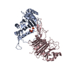
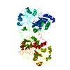
| Structure viewer | Molecule: MolmilJmol/JSmol |
|---|
- Downloads & links
-Download
| PDBx/mmCIF format | 1c6v.cif.gz | 135.5 KB | Display | PDBx/mmCIF format |
|---|---|---|---|---|
| PDB format | pdb1c6v.ent.gz | 105.2 KB | Display | PDB format |
| PDBx/mmJSON format | 1c6v.json.gz | Tree view | PDBx/mmJSON format | |
| Others |  Other downloads Other downloads |
-Validation report
| Arichive directory | https://data.pdbj.org/pub/pdb/validation_reports/c6/1c6vftp://data.pdbj.org/pub/pdb/validation_reports/c6/1c6v | HTTPS FTP |
|---|
-Related structure data


-Links
 PDBj
PDBj- Assembly
| Deposited unit | 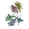
| ||||||||
|---|---|---|---|---|---|---|---|---|---|
| 1 |
| ||||||||
| Unit cell |
| ||||||||
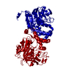
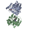
| Details | THERE ARE FOUR CORE DOMAINS AND ONE DNA BINDING DOMAIN IN THE ASYMMETRIC UNIT. THE FOUR CORE DOMAINS ARE LABELLED A A, B, C AND D. THE DNA BINDING DOMAIN ARE LABELLED AS X. THE MISSING RESIDUES ARE: CORE DOMAIN A, A50-A54, A141-A151 CORE DOMAIN B; B50-B54, B141-B151, B208-B212. CORE DOMAIN C; C50-C54, C141-C150, C208-C212. CORE DOMAIN D; D50-D54, D141-D151, C208-C212. DNA BINDING DOMAIN; X214-X215 ,X230-232, X271-X292. |
-Components
| #1: Protein | Mass: 18405.967 Da / Num. of mol.: 4 / Fragment: RESIDUES 813-976 / Mutation: F185H Source method: isolated from a genetically manipulated source Source: (gene. exp.) Simian immunodeficiency virus / Genus: Lentivirus / Plasmid: PET15B / Species (production host): Escherichia coli / Production host:  Escherichia coli K12 (bacteria) / Strain (production host): BL2 / Variant (production host): DE3 / References: UniProt: Q88016 Escherichia coli K12 (bacteria) / Strain (production host): BL2 / Variant (production host): DE3 / References: UniProt: Q88016#2: Protein | | Mass: 9185.444 Da / Num. of mol.: 1 / Fragment: RESIDUES 979-1059 Source method: isolated from a genetically manipulated source Source: (gene. exp.) Simian immunodeficiency virus / Genus: Lentivirus / Plasmid: PET15B / Species (production host): Escherichia coli / Production host: Escherichia coli K12 (bacteria) / Strain (production host): BL2 / Variant (production host): DE3 / References: UniProt: Q87706, UniProt: Q88016*PLUS#3: Water | ChemComp-HOH / | Water Mass: 18.015 Da / Num. of mol.: 66 / Source method: isolated from a natural source / Formula: H2O Mass: 18.015 Da / Num. of mol.: 66 / Source method: isolated from a natural source / Formula: H2O |
|---|
-Experimental details
-Experiment
| Experiment | Method: X-RAY DIFFRACTION |
|---|
- Sample preparation
| Crystal | Density Matthews: 3.61 Å3/Da / Density % sol: 54 % | ||||||||||||||||||||
|---|---|---|---|---|---|---|---|---|---|---|---|---|---|---|---|---|---|---|---|---|---|
| Crystal grow | pH: 5.7 / Details: 0.1 M MES, PH=5.7, PEG6K 8%, 15% DIOXANE | ||||||||||||||||||||
| Crystal grow | *PLUS Method: unknown | ||||||||||||||||||||
| Components of the solutions | *PLUS
|
-Data collection
| Diffraction | Mean temperature: 100 K |
|---|---|
| Diffraction source | Source: SYNCHROTRON / Site: APS  / Beamline: 17-ID / Wavelength: 0.9817 / Beamline: 17-ID / Wavelength: 0.9817 |
| Detector | Type: MARRESEARCH / Detector: CCD / Date: Feb 15, 1999 |
| Radiation | Protocol: SINGLE WAVELENGTH / Monochromatic (M) / Laue (L): M / Scattering type: x-ray |
| Radiation wavelength | Wavelength: 0.9817 Å / Relative weight: 1 |
| Reflection | Resolution: 3→100 Å / Num. obs: 22129 / % possible obs: 92.5 % / Observed criterion σ(I): 3 / Redundancy: 13.9 % / Rmerge(I) obs: 0.113 |
| Reflection shell | Resolution: 3→3.11 Å / Rmerge(I) obs: 0.608 / Mean I/σ(I) obs: 1.5 / % possible all: 66.8 |
| Reflection | *PLUS Num. measured all: 309679 |
| Reflection shell | *PLUS % possible obs: 66.8 % |
- Processing
| Software |
| ||||||||||||||||||||||||||||||||||||||||||||||||||||||||||||
|---|---|---|---|---|---|---|---|---|---|---|---|---|---|---|---|---|---|---|---|---|---|---|---|---|---|---|---|---|---|---|---|---|---|---|---|---|---|---|---|---|---|---|---|---|---|---|---|---|---|---|---|---|---|---|---|---|---|---|---|---|---|
| Refinement | Method to determine structure: SIR, MOLECULAR REPLACEMENT Starting model: PDB ENTRY 1ITG AND 1IHV Resolution: 3→6 Å / Data cutoff high absF: 1000000 / Data cutoff low absF: 0.1 / Cross valid method: THROUGHOUT / σ(F): 2 / σ(I): 2
| ||||||||||||||||||||||||||||||||||||||||||||||||||||||||||||
| Refinement step | Cycle: LAST / Resolution: 3→6 Å
| ||||||||||||||||||||||||||||||||||||||||||||||||||||||||||||
| Refine LS restraints |
| ||||||||||||||||||||||||||||||||||||||||||||||||||||||||||||
| LS refinement shell | Resolution: 3→3.12 Å / Total num. of bins used: 8 /
|
