 Movie
Movie Controller


+ Open data

- Basic information
| Entry | Database: PDB / ID: 1buu | ||||||
|---|---|---|---|---|---|---|---|
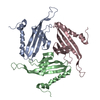
| Title | ONE HO3+ FORM OF RAT MANNOSE-BINDING PROTEIN A | ||||||
 Components Components | PROTEIN (MANNOSE-BINDING PROTEIN A) | ||||||
 Keywords Keywords | SUGAR BINDING PROTEIN /  LECTIN / HOST DEFENSE / METALLOPROTEIN LECTIN / HOST DEFENSE / METALLOPROTEIN | ||||||
| Function / homology |  Function and homology information Function and homology informationcalcium-dependent carbohydrate binding / complement activation, lectin pathway / oligosaccharide binding / killing by host of symbiont cells / collagen trimer / surfactant homeostasis / phosphatidylinositol-4-phosphate binding / polysaccharide binding / protein homotrimerization / D-mannose binding ...calcium-dependent carbohydrate binding / complement activation, lectin pathway / oligosaccharide binding / killing by host of symbiont cells / collagen trimer / surfactant homeostasis / phosphatidylinositol-4-phosphate binding / polysaccharide binding / protein homotrimerization / D-mannose binding / positive regulation of phagocytosis / multivesicular body / complement activation, classical pathway / calcium-dependent protein binding / protease binding / defense response to Gram-positive bacterium / calcium ion binding / protein homodimerization activity / extracellular space / identical protein bindingSimilarity search - Function | ||||||
| Biological species |  Rattus norvegicus (Norway rat) Rattus norvegicus (Norway rat) | ||||||
| Method | X-RAY DIFFRACTION / MOLECULAR REPLACEMENT / Resolution: 1.9 Å | ||||||
 Authors Authors | Ng, K.K.-S. / Park-Snyder, S. / Weis, W.I. | ||||||
 Citation Citation | Journal: Biochemistry / Year: 1998 Title: Ca2+-dependent structural changes in C-type mannose-binding proteins. Authors: Ng, K.K. / Park-Snyder, S. / Weis, W.I. | ||||||
| History |
|
- Structure visualization
| Structure viewer | Molecule: MolmilJmol/JSmol |
|---|
- Downloads & links
-Download
| PDBx/mmCIF format | 1buu.cif.gz | 47.5 KB | Display | PDBx/mmCIF format |
|---|---|---|---|---|
| PDB format | pdb1buu.ent.gz | 32.2 KB | Display | PDB format |
| PDBx/mmJSON format | 1buu.json.gz | Tree view | PDBx/mmJSON format | |
| Others |  Other downloads Other downloads |
-Validation report
| Arichive directory | https://data.pdbj.org/pub/pdb/validation_reports/bu/1buuftp://data.pdbj.org/pub/pdb/validation_reports/bu/1buu | HTTPS FTP |
|---|
-Related structure data
| Related structure data |  1bv4C  1rtmS S: Starting model for refinement C: citing same article ( |
|---|---|
| Similar structure data |


-Links
 PDBj
PDBj


- Assembly
| Deposited unit | 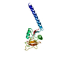
| ||||||||
|---|---|---|---|---|---|---|---|---|---|
| 1 | 
| ||||||||
| 2 | 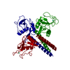
| ||||||||
| Unit cell |
|
-Components
| #1: Protein | Mass: 18449.816 Da / Num. of mol.: 1 Fragment: LECTIN, TRIMERIZATION, AND PORTION OF COLLAGENOUS DOMAIN DOMAINS Source method: isolated from a genetically manipulated source Source: (gene. exp.) Rattus norvegicus (Norway rat) / Organ: LIVER / Plasmid: PINIIIOMPA2 / Cellular location (production host): PERIPLASM / Production host:  Escherichia coli (E. coli) / Strain (production host): JM109 / References: UniProt: P19999 Escherichia coli (E. coli) / Strain (production host): JM109 / References: UniProt: P19999 |
|---|---|
| #2: Chemical | ChemComp-HO /   Mass: 164.930 Da / Num. of mol.: 1 / Source method: obtained synthetically / Formula: Ho Mass: 164.930 Da / Num. of mol.: 1 / Source method: obtained synthetically / Formula: Ho |
| #3: Water | ChemComp-HOH / Water Mass: 18.015 Da / Num. of mol.: 171 / Source method: isolated from a natural source / Formula: H2O Mass: 18.015 Da / Num. of mol.: 171 / Source method: isolated from a natural source / Formula: H2O |
| Sequence details | THE FIRST 4 RESIDUES IN THE EXPRESSION CONSTRUCT, ALA-ILE-GLU-VAL, ARE A CLONING ARTIFACT AND ARE ...THE FIRST 4 RESIDUES IN THE EXPRESSION |
-Experimental details
-Experiment
| Experiment | Method: X-RAY DIFFRACTION / Number of used crystals: 1 |
|---|
- Sample preparation
| Crystal | Density Matthews: 3.8 Å3/Da / Density % sol: 67 % | |||||||||||||||||||||||||||||||||||||||||||||||||
|---|---|---|---|---|---|---|---|---|---|---|---|---|---|---|---|---|---|---|---|---|---|---|---|---|---|---|---|---|---|---|---|---|---|---|---|---|---|---|---|---|---|---|---|---|---|---|---|---|---|---|
| Crystal grow | Method: vapor diffusion, hanging drop / pH: 8 Details: HANGING-DROP VAPOR DIFFUSION AGAINST RESERVOIR CONTAINING 11-15% (W/V) PEG 3350, 0.325 MM HOCL3, 100 MM TRIS-CL PH 8.0, 10 MM NACL, 0.02% NAN3., vapor diffusion - hanging drop | |||||||||||||||||||||||||||||||||||||||||||||||||
| Crystal | *PLUS | |||||||||||||||||||||||||||||||||||||||||||||||||
| Crystal grow | *PLUS | |||||||||||||||||||||||||||||||||||||||||||||||||
| Components of the solutions | *PLUS
|
-Data collection
| Diffraction | Mean temperature: 100 K |
|---|---|
| Diffraction source | Source: ROTATING ANODE / Type: RIGAKU RU200 / Wavelength: 1.5418 |
| Detector | Type: RIGAKU / Detector: IMAGE PLATE / Date: May 15, 1994 / Details: 0.3 MM COLLIMATOR |
| Radiation | Monochromator: GRAPHITE / Protocol: SINGLE WAVELENGTH / Monochromatic (M) / Laue (L): M / Scattering type: x-ray |
| Radiation wavelength | Wavelength: 1.5418 Å / Relative weight: 1 |
| Reflection | Resolution: 1.9→50 Å / Num. obs: 20530 / % possible obs: 95.1 % / Observed criterion σ(I): -3 / Redundancy: 7 % / Biso Wilson estimate: 12.1 Å2 / Rmerge(I) obs: 0.056 / Rsym value: 0.056 |
| Reflection shell | Resolution: 1.9→1.97 Å / Redundancy: 3.9 % / Rmerge(I) obs: 0.215 / Rsym value: 0.215 / % possible all: 88 |
| Reflection shell | *PLUS % possible obs: 88 % |
- Processing
| Software |
| ||||||||||||||||||||||||||||||||||||||||||||||||||||||||||||||||||||||||||||||||
|---|---|---|---|---|---|---|---|---|---|---|---|---|---|---|---|---|---|---|---|---|---|---|---|---|---|---|---|---|---|---|---|---|---|---|---|---|---|---|---|---|---|---|---|---|---|---|---|---|---|---|---|---|---|---|---|---|---|---|---|---|---|---|---|---|---|---|---|---|---|---|---|---|---|---|---|---|---|---|---|---|---|
| Refinement | Method to determine structure: MOLECULAR REPLACEMENT Starting model: PDB ENTRY 1RTM, CHAIN 1 Resolution: 1.9→50 Å / Rfactor Rfree error: 0.005 / Data cutoff high rms absF: 1000000 / Isotropic thermal model: RESTRAINED / Cross valid method: THROUGHOUT / σ(F): 0 Details: MLF TARGET USED. THE FIRST 19 RESIDUES OF THE EXPRESSION CONSTRUCT, CONTAINING SEQUENCE FROM THE COLLAGENOUS DOMAIN, ARE DISORDERED AND CANNOT BE DISTINGUISHED IN ELECTRON DENSITY.
| ||||||||||||||||||||||||||||||||||||||||||||||||||||||||||||||||||||||||||||||||
| Solvent computation | Solvent model: FLAT MODEL / Bsol: 80.9 Å2 / ksol: 0.45 e/Å3 | ||||||||||||||||||||||||||||||||||||||||||||||||||||||||||||||||||||||||||||||||
| Displacement parameters | Biso mean: 28.9 Å2 | ||||||||||||||||||||||||||||||||||||||||||||||||||||||||||||||||||||||||||||||||
| Refine analyze |
| ||||||||||||||||||||||||||||||||||||||||||||||||||||||||||||||||||||||||||||||||
| Refinement step | Cycle: LAST / Resolution: 1.9→50 Å
| ||||||||||||||||||||||||||||||||||||||||||||||||||||||||||||||||||||||||||||||||
| Refine LS restraints |
| ||||||||||||||||||||||||||||||||||||||||||||||||||||||||||||||||||||||||||||||||
| LS refinement shell | Resolution: 1.9→1.97 Å / Rfactor Rfree error: 0.02 / Total num. of bins used: 10
| ||||||||||||||||||||||||||||||||||||||||||||||||||||||||||||||||||||||||||||||||
| Xplor file |
| ||||||||||||||||||||||||||||||||||||||||||||||||||||||||||||||||||||||||||||||||
| Software | *PLUS Name: CNS / Version: 0.3C / Classification: refinement | ||||||||||||||||||||||||||||||||||||||||||||||||||||||||||||||||||||||||||||||||
| Refinement | *PLUS Lowest resolution: 50 Å / σ(F): 0 / % reflection Rfree: 9 % | ||||||||||||||||||||||||||||||||||||||||||||||||||||||||||||||||||||||||||||||||
| Solvent computation | *PLUS | ||||||||||||||||||||||||||||||||||||||||||||||||||||||||||||||||||||||||||||||||
| Displacement parameters | *PLUS Biso mean: 28.9 Å2 | ||||||||||||||||||||||||||||||||||||||||||||||||||||||||||||||||||||||||||||||||
| Refine LS restraints | *PLUS
| ||||||||||||||||||||||||||||||||||||||||||||||||||||||||||||||||||||||||||||||||
| LS refinement shell | *PLUS Rfactor Rfree: 0.271 / % reflection Rfree: 5.7 % / Rfactor Rwork: 0.233 / Rfactor obs: 0.233 |
