 Movie
Movie Controller


+ Open data

- Basic information
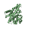
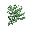
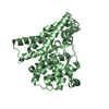
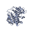
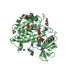
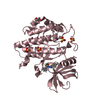
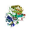
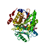
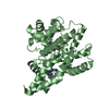
| Entry | Database: PDB / ID: 1boo | ||||||
|---|---|---|---|---|---|---|---|
| Title | PVUII DNA METHYLTRANSFERASE (CYTOSINE-N4-SPECIFIC) | ||||||
 Components Components | PROTEIN (N-4 CYTOSINE-SPECIFIC METHYLTRANSFERASE PVU II) | ||||||
 Keywords Keywords |  TRANSFERASE / TYPE II DNA-(CYTOSINE N4) METHYLTRANSFERASE / AMINO METHYLATION / SELENOMETHIONINE / MULTIWAVELENGTH ANOMALOUS DIFFRACTION TRANSFERASE / TYPE II DNA-(CYTOSINE N4) METHYLTRANSFERASE / AMINO METHYLATION / SELENOMETHIONINE / MULTIWAVELENGTH ANOMALOUS DIFFRACTION | ||||||
| Function / homology |  Function and homology informationsite-specific DNA-methyltransferase (cytosine-N4-specific) / : / site-specific DNA-methyltransferase (cytosine-N4-specific) activity / : / N-methyltransferase activity / DNA restriction-modification system / DNA binding Function and homology informationsite-specific DNA-methyltransferase (cytosine-N4-specific) / : / site-specific DNA-methyltransferase (cytosine-N4-specific) activity / : / N-methyltransferase activity / DNA restriction-modification system / DNA bindingSimilarity search - Function | ||||||
| Biological species |  Proteus vulgaris (bacteria) Proteus vulgaris (bacteria) | ||||||
| Method | X-RAY DIFFRACTION / SYNCHROTRON / MAD / Resolution: 2.8 Å | ||||||
 Authors Authors | Gong, W. / O'Gara, M. / Blumenthal, R.M. / Cheng, X. | ||||||
 Citation Citation | Journal: Nucleic Acids Res. / Year: 1997 Title: Structure of pvu II DNA-(cytosine N4) methyltransferase, an example of domain permutation and protein fold assignment. Authors: Gong, W. / O'Gara, M. / Blumenthal, R.M. / Cheng, X. #1: Journal: Eur.J.Biochem. / Year: 1997Title: Expression, Purification, Mass Spectrometry, Crystallization and Multiwavelength Anomalous Diffraction of Selenomethionyl PvuII DNA Methyltransferase (Cytosine-N4-Specific) Authors: O'Gara, M. / Adams, G.M. / Gong, W. / Kobayashi, R. / Blumenthal, R.M. / Cheng, X. | ||||||
| History |
|
- Structure visualization
| Structure viewer | Molecule: MolmilJmol/JSmol |
|---|
- Downloads & links
-Download
| PDBx/mmCIF format | 1boo.cif.gz | 65.2 KB | Display | PDBx/mmCIF format |
|---|---|---|---|---|
| PDB format | pdb1boo.ent.gz | 50.5 KB | Display | PDB format |
| PDBx/mmJSON format | 1boo.json.gz | Tree view | PDBx/mmJSON format | |
| Others |  Other downloads Other downloads |
-Validation report
| Arichive directory | https://data.pdbj.org/pub/pdb/validation_reports/bo/1booftp://data.pdbj.org/pub/pdb/validation_reports/bo/1boo | HTTPS FTP |
|---|
-Related structure data
| Similar structure data |
|---|

-Links
 PDBj
PDBj- Assembly
| Deposited unit | 
| ||||||||
|---|---|---|---|---|---|---|---|---|---|
| 1 |
| ||||||||
| Unit cell |
| ||||||||
| Noncrystallographic symmetry (NCS) | NCS oper: (Code: given Matrix: (0.6975, -0.6041, 0.3855), Vector : |
-Components
| #1: Protein | Mass: 36956.207 Da / Num. of mol.: 1 Fragment: STARTING FROM THE INTERNAL TRANSLATION INITIATOR AT MET14 Mutation: N TERMIANL DELETION (RESIDUES 1-13) Source method: isolated from a genetically manipulated source Source: (gene. exp.) Proteus vulgaris (bacteria) / Species (production host): Escherichia coliProduction host: Escherichia coli str. K-12 substr. DH10B (bacteria)Strain (production host): DH10B References: UniProt: P11409, site-specific DNA-methyltransferase (cytosine-N4-specific) |
|---|---|
| #2: Chemical | ChemComp-SAH / S-Adenosyl-L-homocysteine  Type: L-peptide linking / Mass: 384.411 Da / Num. of mol.: 1 / Source method: obtained synthetically / Formula: C14H20N6O5S Type: L-peptide linking / Mass: 384.411 Da / Num. of mol.: 1 / Source method: obtained synthetically / Formula: C14H20N6O5S |
-Experimental details
-Experiment
| Experiment | Method: X-RAY DIFFRACTION / Number of used crystals: 2 |
|---|
- Sample preparation
| Crystal | Density Matthews: 4.15 Å3/Da / Density % sol: 38.2 % | ||||||||||||||||||||
|---|---|---|---|---|---|---|---|---|---|---|---|---|---|---|---|---|---|---|---|---|---|
| Crystal grow | pH: 7.2 Details: 0.1 M HEPES PH 7.2, 0.2 M SODIUM ACETATE 20% POLYETHYLENE GLYCOL 400 | ||||||||||||||||||||
| Crystal grow | *PLUS Method: unknown | ||||||||||||||||||||
| Components of the solutions | *PLUS
|
-Data collection
| Diffraction | Mean temperature: 289 K | |||||||||||||||
|---|---|---|---|---|---|---|---|---|---|---|---|---|---|---|---|---|
| Diffraction source | Source: SYNCHROTRON / Site: NSLS  / Beamline: X12C / Wavelength: 0.98233,0.98211,0.92,1.072 / Beamline: X12C / Wavelength: 0.98233,0.98211,0.92,1.072 | |||||||||||||||
| Detector | Type: MARRESEARCH / Detector: IMAGE PLATE / Details: MIRRORS | |||||||||||||||
| Radiation | Protocol: MAD / Monochromatic (M) / Laue (L): M / Scattering type: x-ray | |||||||||||||||
| Radiation wavelength |
| |||||||||||||||
| Reflection | Resolution: 2.8→30 Å / Num. obs: 14886 / % possible obs: 99.7 % / Observed criterion σ(I): 0 / Redundancy: 3.68 % / Rmerge(I) obs: 0.052 / Net I/σ(I): 11.5 | |||||||||||||||
| Reflection shell | Resolution: 2.8→2.85 Å / Rmerge(I) obs: 0.226 / Mean I/σ(I) obs: 5.5 / % possible all: 97.5 | |||||||||||||||
| Reflection | *PLUS Num. measured all: 54787 | |||||||||||||||
| Reflection shell | *PLUS % possible obs: 97.5 % |
- Processing
| Software |
| ||||||||||||||||||||||||||||||||||||||||||||||||||||||||||||
|---|---|---|---|---|---|---|---|---|---|---|---|---|---|---|---|---|---|---|---|---|---|---|---|---|---|---|---|---|---|---|---|---|---|---|---|---|---|---|---|---|---|---|---|---|---|---|---|---|---|---|---|---|---|---|---|---|---|---|---|---|---|
| Refinement | Method to determine structure: MAD / Resolution: 2.8→30 Å / σ(F): 0
| ||||||||||||||||||||||||||||||||||||||||||||||||||||||||||||
| Refinement step | Cycle: LAST / Resolution: 2.8→30 Å
| ||||||||||||||||||||||||||||||||||||||||||||||||||||||||||||
| Refine LS restraints |
| ||||||||||||||||||||||||||||||||||||||||||||||||||||||||||||
| Xplor file | Serial no: 1 / Param file: PARHCSDX.PRO / Topol file: TOPHCSDX.PRO | ||||||||||||||||||||||||||||||||||||||||||||||||||||||||||||
| Software | *PLUS Name: X-PLOR / Version: 3.851 / Classification: refinement | ||||||||||||||||||||||||||||||||||||||||||||||||||||||||||||
| Refine LS restraints | *PLUS
|
